


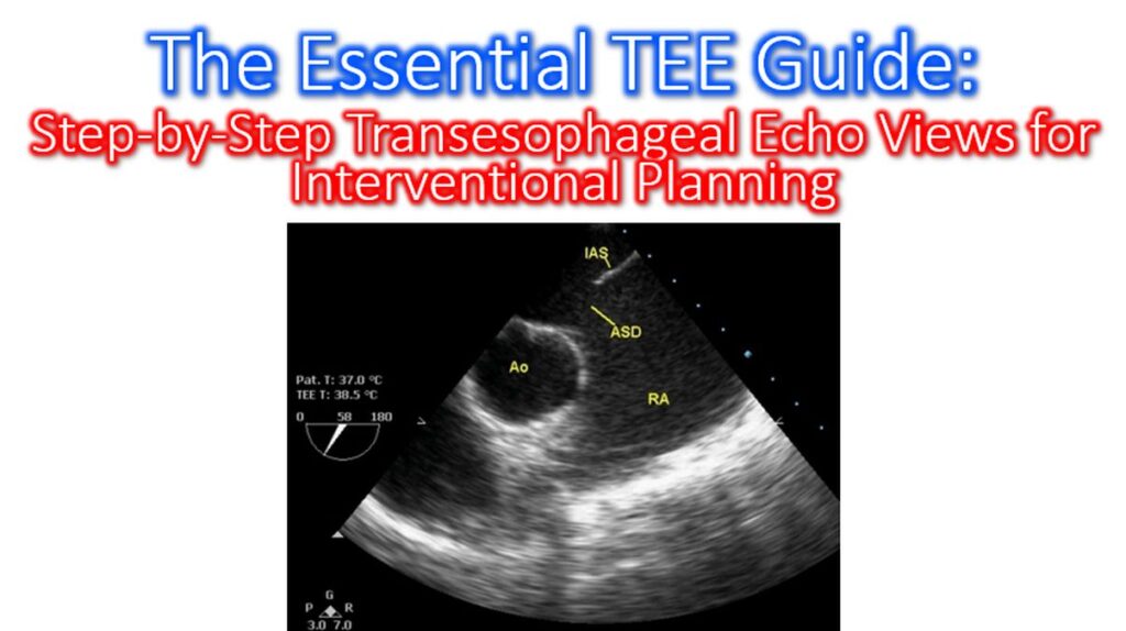

Transesophageal Echocardiography (TEE) is the gold standard for intraprocedural guidance during structural heart interventions. Because it provides a posterior window to the heart, it offers superior resolution of the valves and atrial septal anatomy compared to transthoracic imaging. Here is a step-by-step guide to the essential TEE views required for comprehensive interventional planning.
1. Mid-Esophageal (ME) Four-Chamber View
Angle: Approximately 0° to 20°
- Anatomy: Visualizes the mitral valve (MV), tricuspid valve (TV), and both ventricles.
- Interventional Use: Initial assessment of MV leaflets (A3/P3 and A2/P2) and identifying pericardial effusion. It is the starting point for evaluating right heart function before tricuspid interventions.
2. Mid-Esophageal Mitral Commissural View
Angle: Approximately 60° to 75°
- Anatomy: Visualizes the MV from commissure to commissure (P3–A2–P1).
- Interventional Use: Critical for MitraClip (TEER) procedures. It confirms the clip’s medial-lateral position and ensures the device is centered over the regurgitant jet.
3. Mid-Esophageal Bicaval View
Angle: Approximately 90° to 110°
- Anatomy: Displays the Superior Vena Cava (SVC) on the right and Inferior Vena Cava (IVC) on the left, with the Left Atrium (LA) and Right Atrium (RA) separated by the Interatrial Septum (IAS).
- Interventional Use: The primary view for Transseptal Puncture. It allows the operator to determine the superior-inferior position of the needle on the septum.
4. Mid-Esophageal Long Axis View (LAX)
Angle: Approximately 120° to 140°
- Anatomy: Visualizes the LV outflow tract (LVOT), Aortic Valve (AV), and the A2/P2 segments of the Mitral Valve.
- Interventional Use: Essential for TAVI/TAVR planning to measure the aortic annulus and for gauging the “tenting height” during transseptal punctures for MV repairs.
5. Transgastric Short Axis (SAX) View
Angle: 0° (Advance probe into the stomach and anteflex)
- Anatomy: A “doughnut” view of the Left Ventricle in cross-section.
- Interventional Use: Used to monitor global LV function and regional wall motion during high-risk procedures. It also provides an excellent “en face” view of the mitral valve orifice to check for stenosis post-repair.
Comparison of Key Procedural Views
| Procedure | Primary View | Why? |
| LAA Occlusion | ME 45°, 90°, 135° | Full sweep to measure LAA ostium and depth. |
| ASD/PFO Closure | ME Bicaval (90°) | Visualizes the septal defect and rim adequacy. |
| MitraClip | ME Commissural (60°) | Ensures clip alignment perpendicular to the coaptation line. |
| TAVR | ME LAX (120°) | Precise visualization of the aortic root and valve deployment. |
Clinical Tip: Always perform a 3D en face reconstruction of the Mitral Valve (the “Surgeon’s View”) after obtaining the ME 0° view. This provides the most intuitive map for identifying specific leaflet pathology like prolapse or flail segments. There is a good library of echo clips at University of Toronto website, which includes these five view and other standard TEE views.