


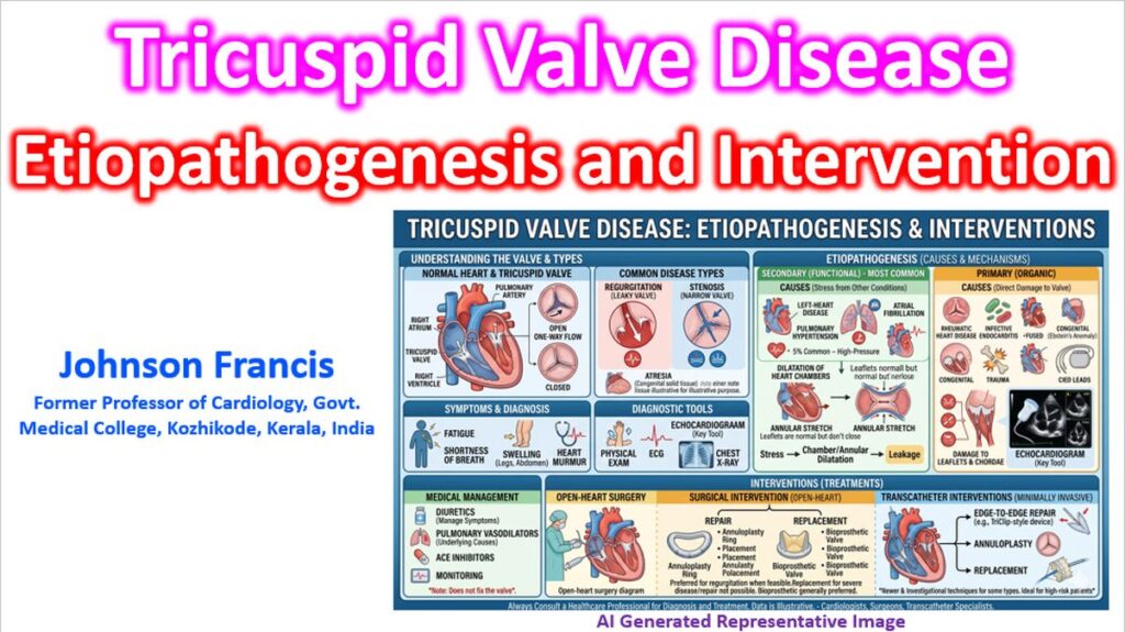

Historically designated as the “forgotten valve,” the tricuspid valve (TV) has undergone a major clinical paradigm shift. Driven by advances in multimodality imaging and transcatheter edge-to-edge repair (TEER), contemporary classification schemas have evolved beyond a simple binary view of tricuspid valve disease (TVD). Understanding its etiopathogenesis requires separating Primary (Organic) disease from Secondary (Functional) phenotypes, the latter of which comprises the vast majority of clinical cases.
1. Secondary (Functional) Tricuspid Regurgitation (FTR)
Secondary TR accounts for over 90% of clinically significant cases in developed nations. In these phenotypes, the valve leaflets and chordae are anatomically normal; regurgitation is entirely driven by altered right atrial (RA) or right ventricular (RV) geometry and shifting hemodynamic loading conditions.
Contemporary guidelines and consensus classifications distinguish between two distinct functional phenotypes based on the primary anatomic driver of remodeling:
A. Atrial Secondary TR (Atrial Functional)
- Primary Driver: Right atrial enlargement and contractile dysfunction, typically precipitated by long-standing atrial fibrillation (AF) or heart failure with preserved ejection fraction (HFpEF).
- Pathomechanism: RA dilation leads to direct expansion and distortion of the tricuspid annulus. Because the leaflets fail to grow proportionally to match the expanding annular area, an imbalance between the leaflet tissue area and the annular orifice develops, resulting in malcoaptation.
- Geometric Features: Minimal or absent leaflet tethering; RV geometry, volume, and longitudinal function remain largely preserved in the early to mid-stages of the disease.
B. Ventricular Secondary TR (Ventricular Functional)
- Primary Driver: RV dilation, spherical remodeling, and systolic dysfunction. This is most commonly secondary to left-sided valvular heart disease (e.g., chronic mitral or aortic valve disease), left ventricular failure, or pre-capillary pulmonary hypertension.
- Pathomechanism: As the RV remodels and dilates, the papillary muscles are displaced apically and laterally. This displacement exerts excessive tension on the chordae tendineae, causing severe systolic leaflet tethering (apical tenting) and preventing normal coaptation.
- Geometric Features: Marked RV structural distortion, pronounced leaflet tenting, and variable degrees of annular dilatation.
2. Primary (Organic) Tricuspid Valve Disease
Primary TVD stems from intrinsic structural, mechanical, or infectious damage to the leaflets, chordae tendineae, or papillary muscles themselves. It represents less than 10% of TR cases but is a leading cause of tricuspid stenosis.
- Infective Endocarditis (IE): A classic primary etiology, frequently seen in the context of intravenous drug use (IVDU) or increasingly associated with cardiac implantable electronic devices (CIEDs) like pacemaker or ICD leads. Leads can cause direct physical perforation, laceration, or mechanical restriction/fibrosis of the leaflets.
- Rheumatic Heart Disease (RHD): Though declining in Western nations, RHD remains a major global cause of organic TVD. It typically presents as diffuse fibrous thickening, fusion of the commissures, and shortening of the chordae, often resulting in a mixed phenotype of tricuspid stenosis and TR.
- Congenital Anomalies: The most notable is Ebstein’s anomaly, characterized by the apical displacement of the septal and posterior leaflets into the RV, effectively “atrializing” a portion of the right ventricle. Other rare congenital etiologies include tricuspid atresia and congenital leaflet clefts.
- Carcinoid Heart Disease: A unique paraneoplastic manifestation of neuroendocrine tumors metastatic to the liver. Vasoactive substances (such as serotonin) cause the deposition of dense, pearly-white fibrous plaques on the endocardial surfaces of the valve. This anchors the leaflets in a semi-open position, causing severe, fixed TR and variable TS.
- Iatrogenic / Trauma: Severe structural TR can result from direct chest wall trauma (e.g., deceleration injuries leading to chordal rupture) or endomyocardial biopsy-induced damage during surveillance of cardiac transplants.
3. The Pathophysics of Annular Remodeling
To understand why secondary TR progresses rapidly, the unique asymmetric architecture of the tricuspid annulus must be highlighted:
The Asymmetric Annular Geometry
Unlike the fibrous, symmetrical mitral annulus, the tricuspid annulus is a highly dynamic, compliant, 3D saddle-shaped structure .
When volume or pressure overload triggers annular dilatation, the remodeling is highly asymmetric:
- Septal Sparing: The septal portion of the annulus is anchored to the fibrous skeleton of the heart and remains relatively resistant to dilation.
- Free-Wall Expansion: Dilatation occurs windows-outwards, predominantly along the free-wall attachments of the anterior and posterior leaflets.
- Planar Flattening: As the annulus expands, it loses its normal 3D saddle configuration, transitioning into a flattened, circular planar geometry. This structural distortion severely undermines coaptation and initiates a vicious cycle: TR causes further right-sided volume overload, accelerating annular dilation and worsening the regurgitation.
4. Modern Clinical & Interventional Implications
The etiopathogenic distinction between these phenotypes heavily dictates timing and selection for intervention. The 2025 ESC/EACTS Guidelines on Valvular Heart Disease introduced a major paradigm shift by upgrading transcatheter tricuspid valve therapies (such as transcatheter edge-to-edge repair or replacement) to a mainstream recommendation (Class IIa) for symptomatic, severe secondary TR.
Crucially, this intervention is highly contingent on early phenotypic identification before the patient reaches an irreversible hemodynamic point-of-no-return, defined by severe, irreversible RV dysfunction or fixed, pre-capillary pulmonary hypertension.
References
de Waha S, Noack T, Kister T, Desch S, Marín-Cuartas M, Kiefer P, Borger MA. Transcatheter and surgical management of tricuspid valve disease: multidisciplinary lifetime management considerations. Ann Cardiothorac Surg. 2026 Mar 31;15(2):18.
Vinciguerra M, Sitges M, Luis Pomar J, Romiti S, Domenech-Ximenos B, D’Abramo M, Wretschko E, Miraldi F, Greco E. Functional Tricuspid Regurgitation: Behind the Scenes of a Long-Time Neglected Disease. Front Cardiovasc Med. 2022 Feb 21;9:836441.
Yucel E, Bertrand PB, Churchill JL, Namasivayam M. The tricuspid valve in review: anatomy, pathophysiology and echocardiographic assessment with focus on functional tricuspid regurgitation. J Thorac Dis. 2020 May;12(5):2945-2954.
Praz F, Borger MA, Lanz J, Marin-Cuartas M, Abreu A, Adamo M et al. 2025 ESC/EACTS Guidelines for the management of valvular heart disease. Eur Heart J 2025;46:4635–736.