Vegetation on aortic valve: Echocardiogram with video narration
Vegetation on aortic valve: Echocardiogram with video narration
Echocardiogram video with narration, showing vegetation on aortic valve
Echocardiogram video in parasternal long axis view demonstrating the large vegetation on the aortic valve prolapsing into the left ventricular outflow tract in diastole and moving outward in systole along with ventricular ejection. The anterior mitral leaflet can be seen below the vegetation, forming the posterior boundary of the left ventricular outflow tract. A scallop of the anterior mitral leaflet can be seen prolapsing into the left atrium in systole. Left atrium is dilated due to the associated mitral regurgitation. Left ventricle is dilated due to both aortic and mitral regurgitation. Both mitral and aortic leaflets are opening well indicating the absence of significant mitral or aortic stenosis. When large vegetations are seen, staphylococcal endocarditis and fungal endocarditis have to be considered. Aortic valve endocarditis is more likely to be acute endocarditis and hence staphylococcal in etiology.
Echocardiogram in parasternal long axis view showing vegetation on aortic valve
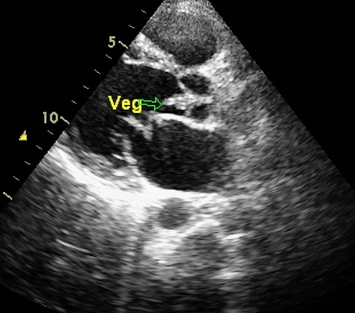
Still frame of two dimensional echocardiogram in parasternal long axis view showing a large vegetation on aortic valve. Aortic valve is in the closed position with part of the vegetation prolapsing into the left ventricular outflow tract (arrow). Below the vegetation, the anterior mitral leaflet is seen, part of which is buckling into the left atrium. The echolucent region below the anterior mitral leaflet is the left atrium and that in front of the mitral valve is the left ventricle. Right ventricular outflow tract region is seen near the apex of the sector near the anterior chest wall. Below the left atrium two circular shadows are seen, one the cross section of the coronary sinus (smaller, upper and to the right of the image) and larger and less defined, below and to the left is the cross section of the descending aorta.
About The Author
Johnson Francis
Former Professor of Cardiology, Calicut Govt. Medical Kozhikode, Kerala, India. Editor-in-Chief, BMH Medical Journal