


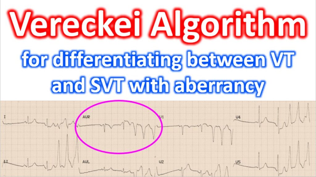

The Vereckei Algorithm is a specific diagnostic protocol used in electrocardiography (ECG) to differentiate between Ventricular Tachycardia (VT) and Supraventricular Tachycardia (SVT) with aberrancy. It was developed as a simpler, often more accurate alternative to the traditional Brugada criteria. It specifically focuses on the morphology of the aVR lead, which is often overlooked in standard rhythm analysis.
The Four Steps of the Algorithm
The algorithm is designed to be followed sequentially. If any step is met, the diagnosis is VT, and you stop. If not, you proceed to the next step.
1. Initial R Wave in aVR
Look at the very beginning of the QRS complex in lead aVR. If there is an initial R wave (a positive deflection right at the start), it is VT.
- Rationale: In SVT with aberrancy, the impulse typically travels away from aVR, leading to an initial Q wave. A primary impulse starting in the ventricles often moves toward aVR. You may recall that aVR is towards the right shoulder and ventricles lower down.
2. Initial r or q wave > 40ms
If there is an initial small r or q wave, measure its width. If it is wider than 40ms (one small box), it is VT.
- Rationale: Ventricular-origin beats move slowly through the myocardium initially, creating a wide initial deflection compared to the rapid conduction of the His-Purkinje system.
3. Notching on the Initial Descending Limb
Look at the downward stroke of a predominantly negative QRS in aVR. If there is notching (a “jagged” or “shaggy” appearance) on that initial descent, it is VT.
4. Ventricular Activation Velocity Ratio (Vi/Vt ≤ 1)
This is the most technical step. You compare the voltage change (ventricular activation-velocity ratio) during the first 40ms (Vi) and the last 40ms (Vt) of the QRS complex. That will correspond to the ratio of the vertical excursion (in millivolts) recorded during the initial and terminal 40 ms of the QRS complex.
- If the initial ventricular activation-velocity is less than or equal to the terminal velocity (Vi /Vt ≤ 1), it is VT.
- If the initial ventricular activation-velocity is faster than the terminal velocity (Vi /Vt > 1), it is SVT.
Why use Vereckei over Brugada?
While both are highly effective, many clinicians prefer the Vereckei method for a few reasons:
- Single Lead Focus: You only need to look at lead aVR, whereas Brugada requires looking across all precordial leads (V1 through V6).
- Speed: In emergency settings (like an ER or CCU), analyzing one lead is often faster.
- Accuracy: Some studies suggest the “aVR-only” approach has higher specificity for identifying VT in patients with pre-existing bundle branch blocks.
Clinical Pearl
Always remember the “Golden Rule” of ACLS: If the patient is unstable and has a wide-complex tachycardia, treat it as VT regardless of the algorithm. These tools are best utilized when the patient is hemodynamically stable and you have the time for a detailed 12-lead analysis.
The original reference for the Vereckei Algorithm actually consists of two primary papers. While the term is most commonly associated with the 2008 “aVR-only” version, it was preceded by a 2007 multistep version.
1. The aVR-only Algorithm (Most Common)
This is the four-step protocol focusing exclusively on lead aVR that is widely taught today.
- Full Citation: Vereckei A, Duray G, Szénási G, Altemose GT, Miller JM. New algorithm using only lead aVR for differential diagnosis of wide QRS complex tachycardia. Heart Rhythm. 2008 Jan;5(1):89-98.
- Key Finding: This study demonstrated that a single-lead (aVR) approach was superior in accuracy to the Brugada criteria, primarily because it eliminated complex morphological interpretations in the precordial leads.
2. The Original Multistep Algorithm
Before the aVR-only version, Vereckei and colleagues published a broader algorithm that included AV dissociation and other morphological features across the 12-lead ECG.
- Full Citation: Vereckei A, Duray G, Szénási G, Altemose GT, Miller JM. Application of a new algorithm in the differential diagnosis of wide QRS complex tachycardia. European Heart Journal. 2007 Mar;28(5):589-600.
- Key Finding: This paper introduced the Vi/Vt ratio (Ventricular Activation Velocity Ratio) as a concept, which later became Step 4 of the 2008 aVR-only algorithm.
Comparison of the Two Papers
| Feature | 2007 Paper (Original) | 2008 Paper (Simplified) |
| Leads Used | Full 12-lead ECG | Lead aVR only |
| Step 1 | Search for AV Dissociation | Initial R wave in aVR |
| Step 4 | Vi/Vt ratio in any lead | Vi/Vt ratio in aVR |
| Primary Goal | Improve Brugada criteria | Simplify to a single lead |
If you are citing this for clinical guidelines or an exam, the 2008 Heart Rhythm paper is usually the one intended when people refer to the “Vereckei aVR Algorithm.”