VVI pacing with ectopic beat and fusion beat
VVI pacing with ectopic beat and fusion beat
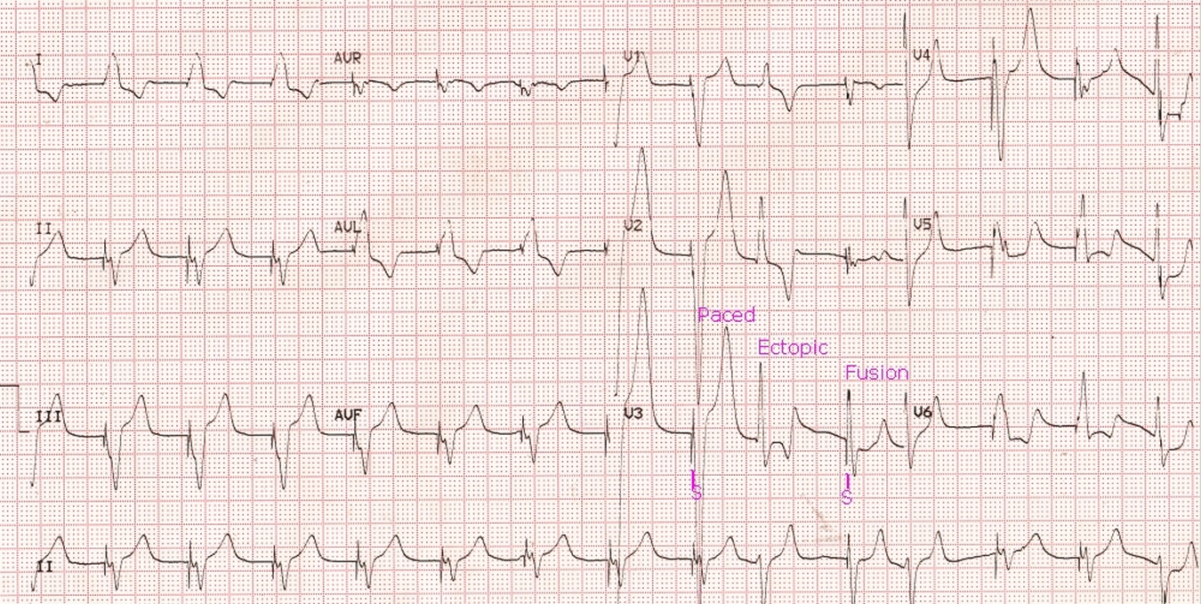 VVI pacing with ectopic beat and fusion beat: Basic rhythm in this ECG is a paced rhythm, with pacing spikes (pacemaker artefacts) seen immediately before each QRS complex. The QRS complexes have a left bundle branch block pattern with a superior axis, indicating right ventricular apical pacing. An ectopic beat with right bundle branch block morphology has occurred in between, causing the pacemaker to be inhibited, indicating that it is a demand pacemaker working in VVI mode. In VVI mode, the chamber paced and sensed is the ventricle and the response to a sensed beat is inhibition of pacing. After the regular pacing cycle, the next spike (S) occurs, but the QRS has a morphology intermediate between the spontaneous ectopic beat and the paced beat, with a slightly narrower QRS than the paced beat. This is a fusion beat between a paced beat and a spontaneous ectopic beat which has occurred just after the onset of the pacing spike. When a fusion beat occurs during a ventricular tachycardia, they are called Dressler beats [1, 2].
The ectopic beat could either have a left ventricular origin due to the RBBB morphology or a supraventricular origin with aberrant conduction. Occasionally, sinus beats can occur during a ventricular tachycardia and are called capture beats. They are recognized by narrow QRS and preceding P waves. Capture beats are more likely to occur in slower ventricular tachycardias.
References
- 1.William Dressler, Hugo Roesler. The occurrence in paroxysmal ventricular tachycardia of ventricular complexes transitional in shape to sinoauricular beats; a diagnostic aid. Am Heart J. 1952 Oct;44(4):485-93.
- Young RL, Mower MM, Ramapuram GM, Tabatznik B. Atrial fibrillation with ventricular tachycardia showing “Dressler” beats. Chest. 1973 Jan;63(1):96-7.
About The Author
Johnson Francis
Former Professor of Cardiology, Calicut Govt. Medical Kozhikode, Kerala, India. Editor-in-Chief, BMH Medical Journal