


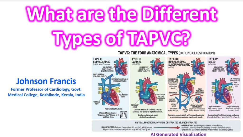

Total Anomalous Pulmonary Venous Connection (TAPVC)—also referred to as TAPVR (Return)—is universally categorized using the Darling Classification, which groups the anomaly based on the anatomical site where the pulmonary veins drain into the systemic venous circulation. Because the pulmonary veins fail to connect to the left atrium, oxygenated blood mixes entirely in the right atrium. Regardless of the type, an obligatory right-to-left shunt (an unrestrictive ASD or PFO) is required to sustain life.
The Four Anatomical Types
1. Type I: Supracardiac (Most Common, ~50%)
The four pulmonary veins gather into a common posterior venous confluence behind the left atrium.
- The Pathway: Blood drains upward from this confluence into an ascending vertical vein (a remnant of the embryonic left superior vena cava), which empties into the left brachiocephalic (innominate) vein, flows into the right superior vena cava (SVC), and finally enters the right atrium.
- Classic Sign: This creates the classic widened mediastinum known as the “Snowman sign” (or figure-of-8) on a frontal chest X-ray in older infants.
2. Type II: Cardiac (~20–25%)
The pulmonary veins drain directly into the heart itself, bypassing the systemic veins entirely.
- The Pathway: The posterior confluence connects directly to the Coronary Sinus (which becomes massively dilated) or, less commonly, opens directly into the posterior wall of the Right Atrium.
- Clinical Nuance: This type is usually unobstructed and carries the most straightforward surgical repair (unroofing the coronary sinus into the left atrium and baffling the ASD).
3. Type III: Infracardiac / Subdiaphragmatic (~20%)
The pulmonary veins drain downward below the diaphragm.
- The Pathway: The confluence empties into a descending vertical vein that passes through the esophageal hiatus of the diaphragm. It typically connects to the Portal Vein, the Ductus Venosus, the Hepatic Veins, or the Inferior Vena Cava (IVC).
- Clinical Nuance: This is a true pediatric cardiac emergency. It is almost universally obstructed because the vertical vein is squeezed as it passes through the diaphragm, or the drainage gets choked off when the ductus venosus naturally closes postnatally. Neonates present rapidly with profound cyanosis, severe pulmonary edema, and cardiogenic shock.
4. Type IV: Mixed (~5–10%)
A combination of two or more of the above drainage pathways.
- The Pathway: The most frequent combination involves the left pulmonary veins draining via a supracardiac vertical vein, while the right pulmonary veins drain directly into the coronary sinus.
The Critical Functional Division: While Darling’s types dictate the anatomy, the most important clinical classification is Obstructed vs. Unobstructed. An unobstructed supracardiac TAPVC might present at 1 to 2 months of age with mild cyanosis and right-sided volume overload (mimicking a large ASD). Conversely, an obstructed infracardiac TAPVC presents within hours of birth with a “snowstorm” appearance on a chest X-ray that is routinely misdiagnosed as severe Respiratory Distress Syndrome (RDS).