


Mitral regurgitation in PLAX and Apical 4C views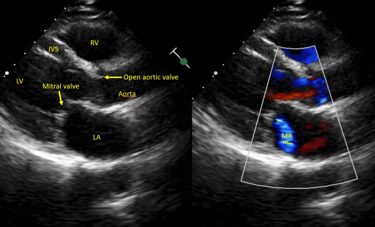

Parasternal long axis view showing mitral regurgitation jet into the left atrium. MR is seen as a bluish tongue shaped mosaic jet directed downwards into the left atrium, in the left panel. Aortic valve is seen as open and the mitral valve seen in closed position as it is a systolic frame.


Caution is needed while quantifying MR jet because the visible area can vary depending on the instrument settings and hemodynamic variables. Acute severe MR with hypotension can have very small jet area while hypertensive patients with mild MR can have large jet area [1]. That is why we always insist for a clinical evaluation prior to echocardiography, which will give a lot of valuable hints. The person with hypertension with mild MR will be clinically quite different from the person with acute severe MR and hypotension. Latter will most likely be in an intensive care unit, quite sick and unlikely to be brought to the echo room, rather we will be doing a bedside study!
Reference
- Grayburn PA, Weissman NJ, Zamorano JL. Quantitation of mitral regurgitation. Circulation. 2012 Oct 16;126(16):2005-17.