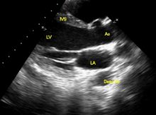
Parasternal long axis view in normal echocardiogram Parasternal long axis view in normal echocardiogram: RV FW: Right ventricular free wall; RV: Right ventricle; IVS: Interventricular septum; Ao: Aorta;
PLAX view in MS Echocardiogram in parasternal long axis view in mitral stenosis. The anterior mitral leaflet is seen to be domed, with a hockey stick appearance. The
Mitral regurgitation in PLAX and Apical 4C views Parasternal long axis view showing mitral regurgitation jet into the left atrium. MR is seen as a bluish tongue shaped
Modified PLAX view in TGA Parasternal long axis (PLAX) view is often the first view taken during echocardiography. Usually it visualizes the left ventricle, left atrium, right ventricular
Echocardiogram in Aortic Stenosis Echocardiogram is a very useful tool to assess aortic stenosis. It will identify whether the stenosis is valvar, supra valvar or subvalvar. Associated lesions
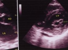
Echocardiogram in Mitral Stenosis Left panel shows the parasternal long axis view (PLAX). AO: Aorta; MVO: Mitral valve opening; LA: left atrium. Doming of the anterior mitral leaflet
In which echocardiographic view was this image recorded? a. Subcostal view
b. Apical four chamber view
c. Parasternal long axis view
d. Parasternal short axis view