Angiography
What is angiography?
Angiography (angio) is visualisation of a blood vessel with or without injecting a contrast (‘dye’) into its lumen. Most of us are now familiar with coronary angiography used to visualise the blood vessels supplying oxygenated blood to the heart. Usually visualization of each blood vessel is named after it: e.g. renal angio – visualisation of renal (kidney) vessels; cerebral angio – visualization of blood vessels of cerebral (brain) vessels; peripheral angio – visualisation of the blood vessels of the limbs and other peripheral organs.
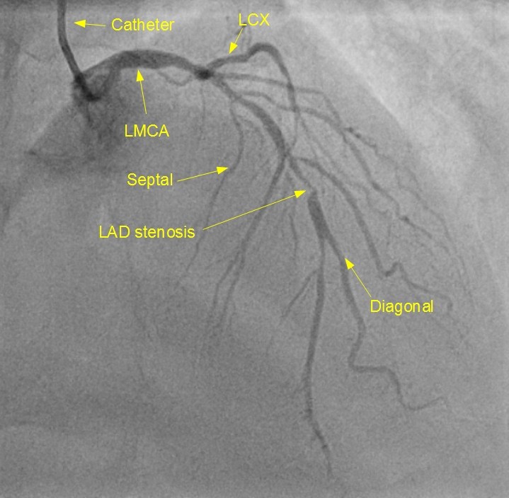
LMCA: Left main coronary artery; LCX: Left circumflex coronary artery; LAD stenosis: Narrowing of left anterior descending (LAD) coronary artery; Septal: Septal branch of left anterior descending coronary artery; Diagonal: Diagonal branch of left anterior descending coronary artery: Catheter: Small tube used to inject radiocontrast dye into the coronary artery for opacification and visualization. This angiographic picture shows the most important left sided vessels supplying oxygenated blood to the heart. Septal branches supply the interventricular septum, the wall between two lower chambers of the heart. Diagonal branches supply the left side of the left ventricle, which is the lower muscular chamber of the heart. Left circumflex artery curves around to the back of the heart to supply that region. Left anterior descending artery supplies the front of the heart. Both are branches of the left main coronary artery.
The right side of the heart is supplied by right coronary artery (RCA) which is not seen in this picture.
What are the different techniques of angiography?
Angiography can be divided into invasive and non invasive depending how much invasion is done into the body structures for visualising the vessels. Magnetic resonance angiography (MR angio) can be considered as the truly non invasive angio as there is no need to inject any contrast into the blood vessel to visualize the vessels. The hydrogen ions in the water content itself acts as the contrast for MR angiography. Hence MR angio is also known as ‘dyeless angiography‘.
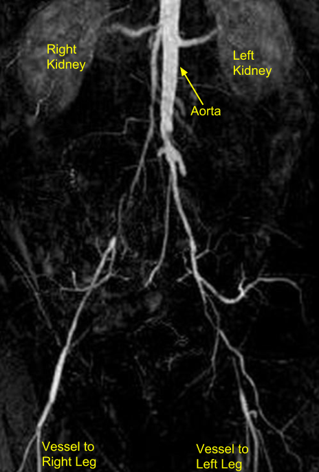
The picture shows magnetic resonance angiogram of the abdominal aorta (large blood vessel inside the tummy). The two bean shaped structures seen on either side in the upper part are the kidneys, with a branch to each arising from the aorta in the middle. In the lower part of the figure vessels going to the lower limbs can be seen. Stumps are noted in lower part of aorta due to obstructed branch vessels.
In computed tomographic (CT) angio, iodinated contrast is injected into the peripheral veins (blood vessels carrying deoxygenated blood to the heart) of the forearms. X-ray equipment then captures the movement of the contrast into various parts of the body. If if captures the coronary blood vessels, then it is called CT coronary angio. If it captures the blood flow to the lungs, it is called CT pulmonary angio.
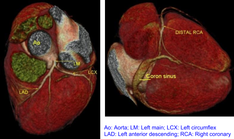
These are three dimensional reconstructions of CT coronary angiograms. Initial image shows the left main coronary artery arising from the aorta and its branches. Second image shows the under surface of the heart with coronaray sinus, the large blood vessel which drains the deoxygenated blood from the heart to the right atrium (upper thin walled chamber).
In truly invasive angio like coronary angio, the contrast is injected directly into the blood vessels of the heart (coronary arteries). This is done by introducing small tubes known as catheters under local anaesthesia through the blood vessels at the wrist or groin and tacking them under X-ray fluoroscopic guidance to the coronary arteries through the aorta (largest blood vessel carrying oxygenated blood, arising from the heart).
What is fluorescein angiography?
Fluorescein angiography is used to visualise the blood vessels of the eye by injecting fluorescent dye into the blood vessels. The images of the inner eye are then photographed, to study the blood vessels of the retina, the light sensitive inner coating of the eye which senses our visual information and transmits it to the brain.
Related Posts


About The Author
Johnson Francis
Former Professor of Cardiology, Calicut Govt. Medical Kozhikode, Kerala, India. Editor-in-Chief, BMH Medical Journal