Coronary CT angiogram
Coronary CT angiogram
Coronary CT angiograms are increasing in popularity as a non-invasive screening tool for detecting significant coronary artery disease. Coronary arteries are blood vessels supplying oxygenated blood to the heart. Angiograms are images of blood vessels, usually obtained by injecting medications for contrast from other body structures.
CT angiograms are reconstructions from 64 or more slice CT scans following injection of radiocontrast dye into a forearm vein. Veins are blood vessels returning deoxygenated blood to the heart. This can be done as an outpatient test, in the X-ray department.
As of now CT angiograms cannot replace conventional coronary angiograms for assessing the finer details of coronary arteries while planning for procedures like balloon angioplasty. Balloon angioplasty is removal of blocks from the blood vessels using slender long tubes with high pressure balloons at the tip.
Conventional or invasive coronary angiograms are obtained by injecting radiocontrast medications and recording continuous images using specialized X-ray equipment in a cardiac catheterization laboratory. The dye is injected directly into the coronary arteries through long tubes known as catheters introduced through blood vessels at the wrist or groin.
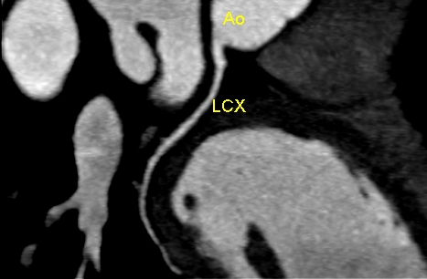
A simple CT image of a blood vessel of the heart will look like this in a two dimensional view. One blood vessel has been marked as LCX and is seen arising from aorta, marked as Ao, which is the largest blood vessel arising from the heart and supplying the whole body.
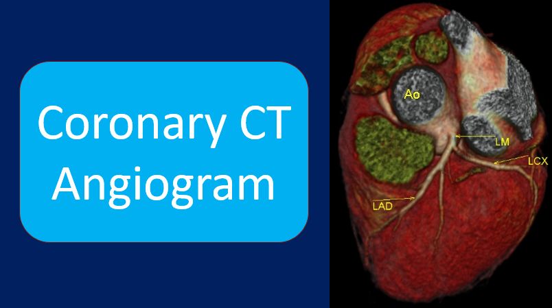
Reconstructed three dimensional image will look like this. Here we can see the left main coronary artery arising from the aorta, and has been marked as LM. Two branches of the left main have been marked LAD and LCX.
These are the two major blood vessels supplying oxygenated blood to the heart. Those of you have had opportunity to read angiogram reports would have noticed these names in the reports.

This is another view, which shows these blood vessels, but left main has been marked as LMCA, short for left main coronary artery. In addition, the major coronary vein known as coronary sinus has also been marked. Coronary sinus drains the deoxygenated blood from the heart.

A view from the right side of the heart shows the right coronary artery which has been marked as RCA. This is another major blood vessel supplying oxygenated blood to the heart. Sudden blockage of any of these blood vessels can cause a heart attack. CT angiogram is useful in detecting major blocks in these blood vessels.

Related Posts


About The Author
Johnson Francis
Former Professor of Cardiology, Calicut Govt. Medical Kozhikode, Kerala, India. Editor-in-Chief, BMH Medical Journal