Device closure of PDA
Device closure of PDA
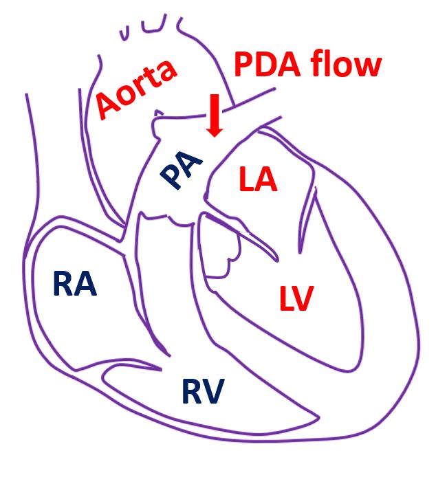
Patent ductus arteriosus known in short as PDA is a birth defect in which a communication between the aorta and pulmonary artery persists after birth. Normally this opening which is present in the baby before birth, closes off soon after birth. When it persists, blood flows from the aorta to the pulmonary artery. Aorta is the largest blood vessel in the body carrying oxygenated blood. Pulmonary artery carries blood to the lungs for oxygenation. When the PDA is large, blood flow to lungs increases very much and pressure in the blood vessels of the lungs increase and can cause heart failure.
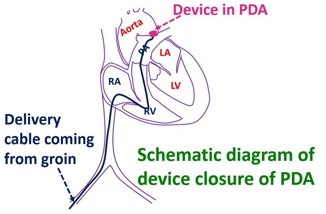
Small PDAs can be closed by coils inserted into them which will promote formation of clot and close off the defect. Large PDAs require a specially made device to close them. These devices folded like an umbrella are mounted on delivery cables and inserted through delivery sheaths. Delivery sheaths are introduced through blood vessels in the groin and guided to the heart by continuous X-ray imaging. The fluoroscopic X-ray picture shows a PDA device in the desired location, still attached to the delivery cable.

The position of the device is checked by X-ray and small amount of radiocontrast medication is injected to ensure that there is no residual leak. Ultrasound study of the heart known as echocardiogram is also useful to ensure correct position of the device and absence of leak. Once everything is fine, the delivery cable is unscrewed and the device released. Delivery cable and sheath are taken out then.
Device closure of PDA can be done under local anaesthesia in older children who are co-operative. In young children who are afraid of the procedure, general anaesthesia is required. As it is not an open surgery, hospital stay needed is usually just one day or even lesser. Long term results are good with device closure of PDA. Very rarely, device can erode into nearby vital structures of the heart. This is mostly avoided by meticulous pre-procedure planning and attention to finer details of the procedure.
Related Posts


About The Author
Johnson Francis
Former Professor of Cardiology, Calicut Govt. Medical Kozhikode, Kerala, India. Editor-in-Chief, BMH Medical Journal