Concerns about radiation exposure in cardiac CT angiography Cardiac CT angiography is a popular test for non-invasive evaluation of coronary artery disease or blocks in the blood vessels
Cardiac Invalidism ‘Cardiac Invalidism’ refers to persons experiencing physical and psychological disability even though they have adequate physical recovery from an episode of Myocardial Infarction or heart attack.
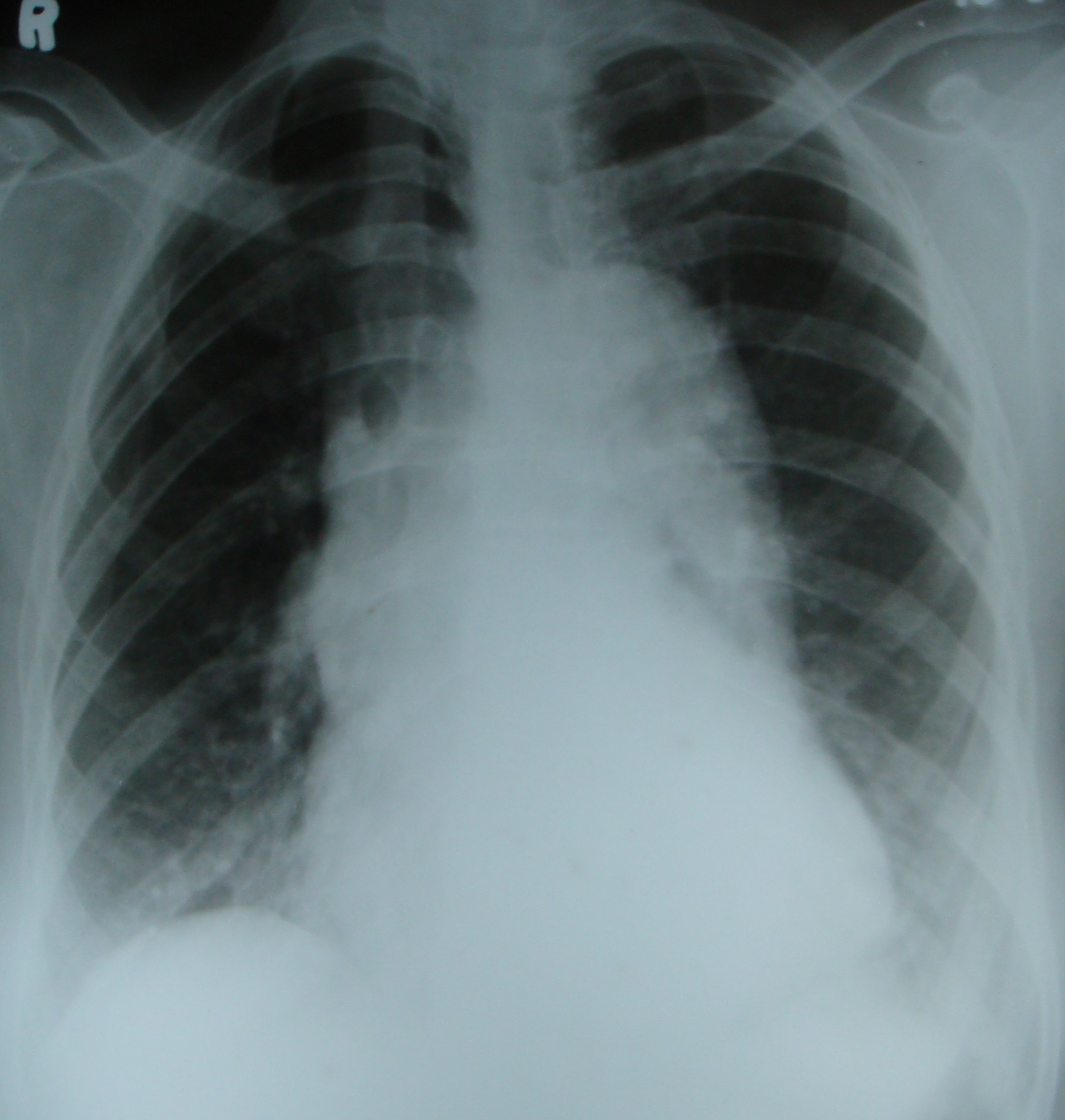
Rotated Chest X-ray Rotated chest x-ray with prominent aorta. Apparent tracheal shift to the right is due to rotation. Medial end of right clavicle is seen farther away
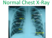
Normal Chest X-Ray Normal chest X-ray, PA view demonstrating cardiovascular structures. RPA: right pulmonary artery; LPA: left pulmonary artery; MPA: main pulmonary artery. Right heart border is formed
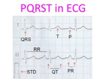
PQRST in ECG The waves and intervals in a normal ECG are illustrated here. P wave is due to atrial depolarisation. QRS is the ventricular depolarisation and T
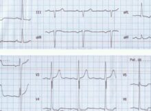
Left ventricular hypertrophy Left ventricular hypertrophy is manifest in this ECG as tall R waves in lateral leads (leads I, aVl, V5 and V6). S wave in V1
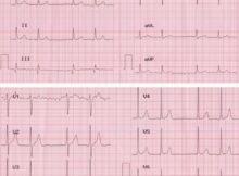
Atrial Fibrillation with Bigeminal Rhythm Baseline is irregular and lead V1 shows coarse fibrillary waves suggestive of atrial fibrillation. But the unusual feature about this ECG is the
Heart disease in mice reversed Tiny fragments of genetic material may play a great role in the development of heart disease. By modifying the genetic material, scientists have
How is embolisation of an artery done? First an angiogram is done by injecting the main artery with iodinated contrast to visualise the origin of the target vessel from