Pulmonary Stenosis
Pulmonary Stenosis
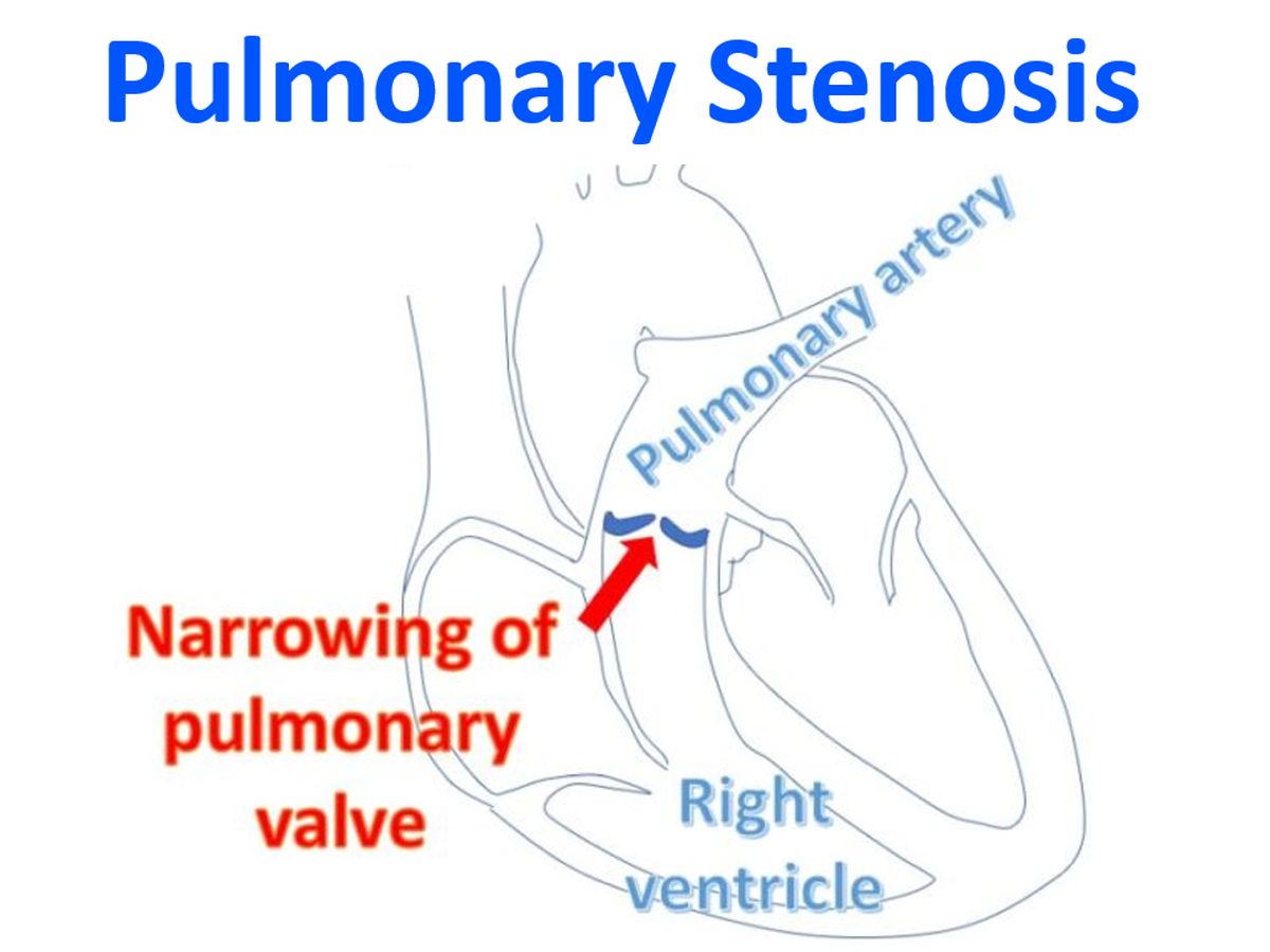
Pulmonary stenosis is a congenital narrowing of the pulmonary valve. Severe PS leads to right ventricular hypertrophy and failure. Right ventricular end diastolic pressure elevation leads to elevated right atrial pressure. Elevated right atrial pressure stretches open the foramen ovale and consequent right to left shunting of blood. This leads to systemic desaturation. PS is a component of many complex congenital heart diseases like Tetralogy of Fallot, the severity of which is determined by the severity of PS.
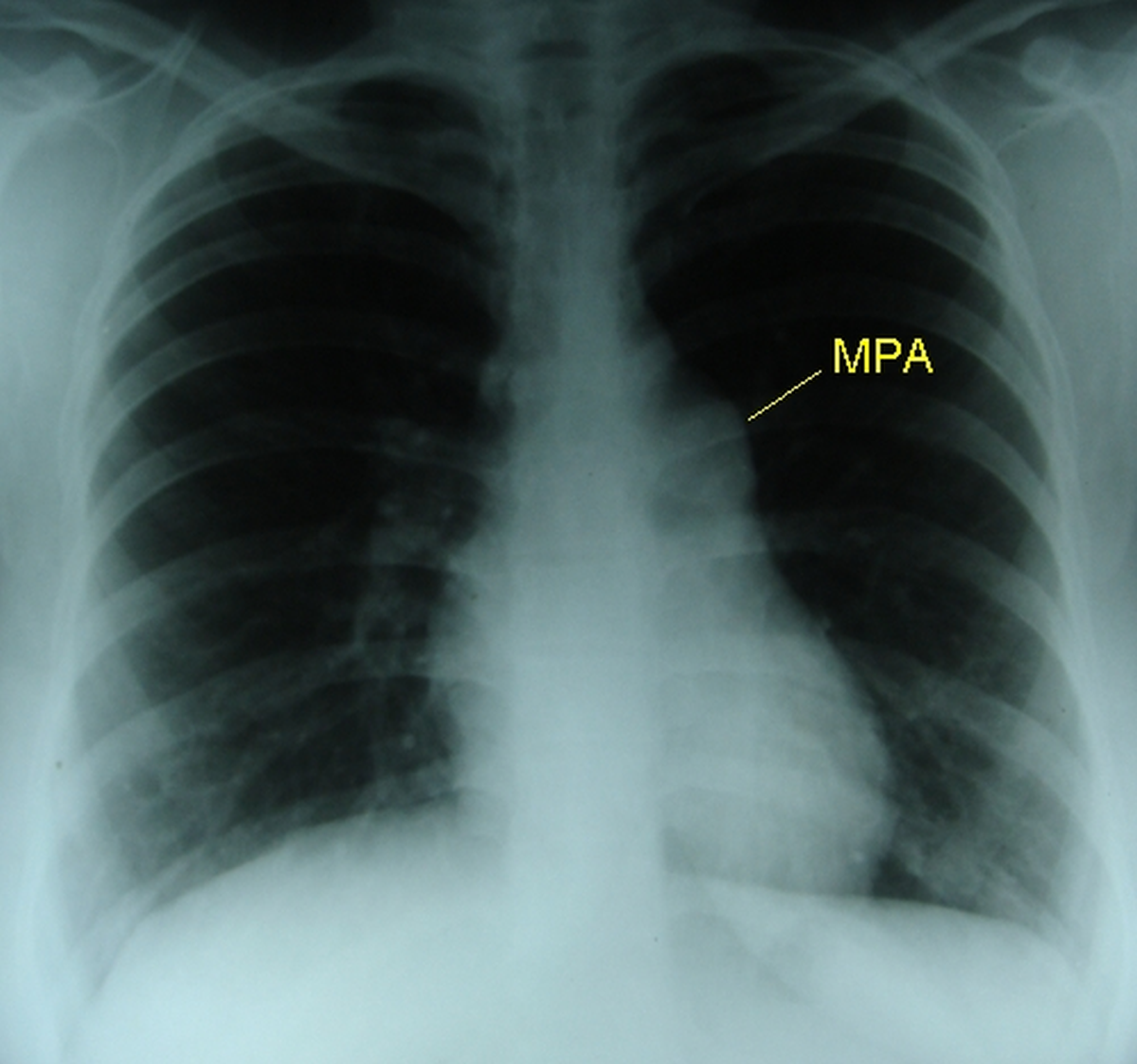
ECG shows right ventricular hypertrophy and strain pattern in severe PS. Chest X-ray shows dilatation of main pulmonary artery (post stenotic dilatation) and left pulmonary artery. Right pulmonary artery is not dilated in pulmonary stenosis. This contrasts the x-ray finding in PS from that in pulmonary hypertension where both left and right pulmonary artery as well as main pulmonary artery are dilated.
Mild and moderate PS will be virtually asymptomatic and well tolerated. They do not require any specific treatment. Severe pulmonary stenosis requires balloon pulmonary valvotomy. Severity can be assessed by Doppler echocardiography and by cardiac catheterisation. Balloon pulmonary valvotomy gives good results and is a relatively low risk procedure, compared to the open pulmonary valvotomy which was being practised earlier.
Related Posts


About The Author
Johnson Francis
Former Professor of Cardiology, Calicut Govt. Medical Kozhikode, Kerala, India. Editor-in-Chief, BMH Medical Journal