Tests for heart disease
What are the important tests for heart disease?
It should be noted that before any tests, a good medical evaluation is needed to decide which tests will be most useful for that particular person. Otherwise it may be a wastage of resources and the needed result may not be obtained. In general, tests are most useful when there is an intermediate probability of disease. When there is a low probability, a positive result could be false positive. Similarly, when the probability is high, a negative result may be a false negative. So we have to know the pretest probability of the disease before proceeding with a test, which is what we plan to achieve by a good medical evaluation.
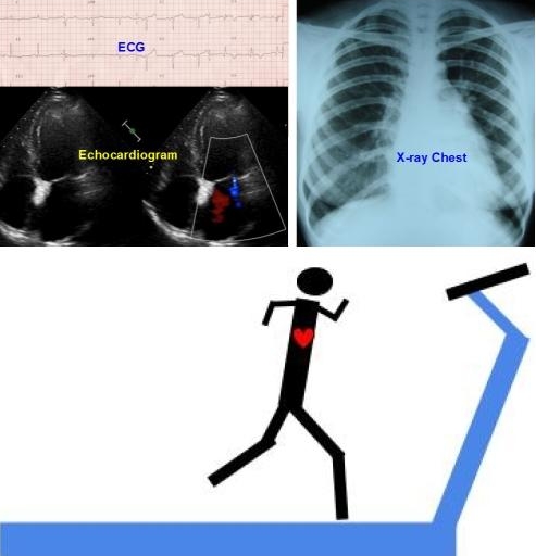
One of the first tests to be done when heart disease is suspected, is an ECG, which records the electrical activity of the heart. Though it was started over a century back, it is still one of the best tests to diagnose a heart attack at the bedside. It does have its limitations – a normal ECG does not rule out heart disease. X-ray of the chest is useful to document the size of the heart and to look for enlargement of various chambers and blood vessels. It is also a simple and old test, with certain advantages and disadvantages.
Blood tests used in the emergency department to detect heart disease are Troponin estimation to detect heart attack and BNP estimation to look for evidence of heart failure.
Next come the imaging tests. Ultrasound study of the heart is known as echocardiography, which can detect the function of the heart and structural abnormalities of the heart, major blood vessels and heart valves. It is now available in portable mode at the bedside. Cardiac computerized tomography or cardiac CT is the next imaging test, which can be done at the radiology suite.
An advanced form of cardiac CT is CT coronary angiography. Magnetic resonance imaging (MRI or CMR – cardiac magnetic resonance imaging) is yet another advanced mode of cardiac imaging. Nuclear scans of the heart include imaging the blood pool with radioactive tracers, imaging the blood flow to the heart with tracers and assessing the metabolic function of the heart with PET scan (positron emission tomography).
Holter monitoring and treadmill tests are different forms of ECG evaluations in different settings. Holter is continuous ambulatory monitoring done for 24-48 hours. Treadmill test is exercise ECG, done with a computerized treadmill which monitors and records the ECG during gradual, stage wise increasing speeds of the treadmill.
Electrophysiological study is an invasive form of ECG in which leads are introduced into the heart through blood vessels and recording of the electrical activity from various sites of the heart done from inside.
Gold standard for diagnosing coronary artery disease is coronary angiography. Allied tests to coronary angiography are coronary intravascular ultrasound (IVUS), fractional flow reserve (FFR) and optical coherence tomography (OCT). All these are invasive tests, requiring the introduction of small tubes known as catheters into the blood vessels and the procedure of introducing catheters into the heart is known as cardiac catheterization. Cardiac catheterization is done in a cardiac catheterization laboratory equipped with cine X-ray equipment.
Related Posts


About The Author
Johnson Francis
Former Professor of Cardiology, Calicut Govt. Medical Kozhikode, Kerala, India. Editor-in-Chief, BMH Medical Journal