What is aortic aneurysm?
What is aortic aneurysm?
Aortic aneurysm is a localized enlargement of the aorta. Aorta is the largest blood vessel which carries oxygenated blood to the body. The aneurysm can increase in size as time progresses and can even rupture occasionally, with catastrophic results. Operations and other procedures done when the aneurysm is enlarging rapidly can prevent this catastrophe.
Risk factors for development of an aortic aneurysm is similar to those for a heart attack, though both are entirely different conditions. Heart attacks are due to blocks in blood vessels of the heart. High blood pressure increases the chance for development of aortic aneurysm. It is also a risk factor for heart attack.
Male gender is another risk factor, with aortic aneurysms being commoner in males than females. Advancing age is another risk factor, more so after the age of 65 years. In fact several countries have implemented mandatory periodic screening for aneurysm of the aorta in the tummy (abdominal aortic aneurysm, AAA) with ultrasound examination.
Smoking is another important risk factor for heart attack and aortic aneurysm. Longer duration and higher usage increases the risk. Obesity is also a risk factor in common for heart attack and aortic aneurysm.
There is also a familial tendency for aortic aneurysm. There are certain genetically transmitted conditions like Marfan syndrome and Ehlers-Danlos syndrome which predispose to aortic aneurysm. In these conditions, there is a defect in the support to the aortic wall due to connective tissue disease.
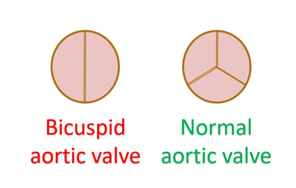
Bicuspid aortic valve is another important association of aortic aneurysm. Normally the aortic valve has three semilunar leaflets. Bicuspid aortic valve is a birth defect in which there are only two leaflets instead of three. It can be associated with disease of the aortic wall known as aortopathy. Those with aortopathy can develop progressive enlargement of the aorta and lead to aneurysm formation.
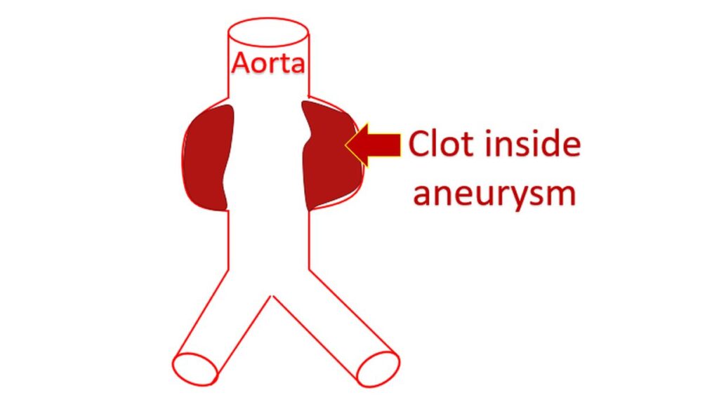
Aortic aneurysm can produce local symptoms depending on the location, due to compression of nearby structures. Important sites for the aneurysm are just beyond the aortic valve, in the chest and in the tummy. Sometimes a clot can form inside the aneurysm. Part of the clot can break away and get carried in the blood circulation. It can get lodged in a branch blood vessel and block it, damaging the organ supplied by it.
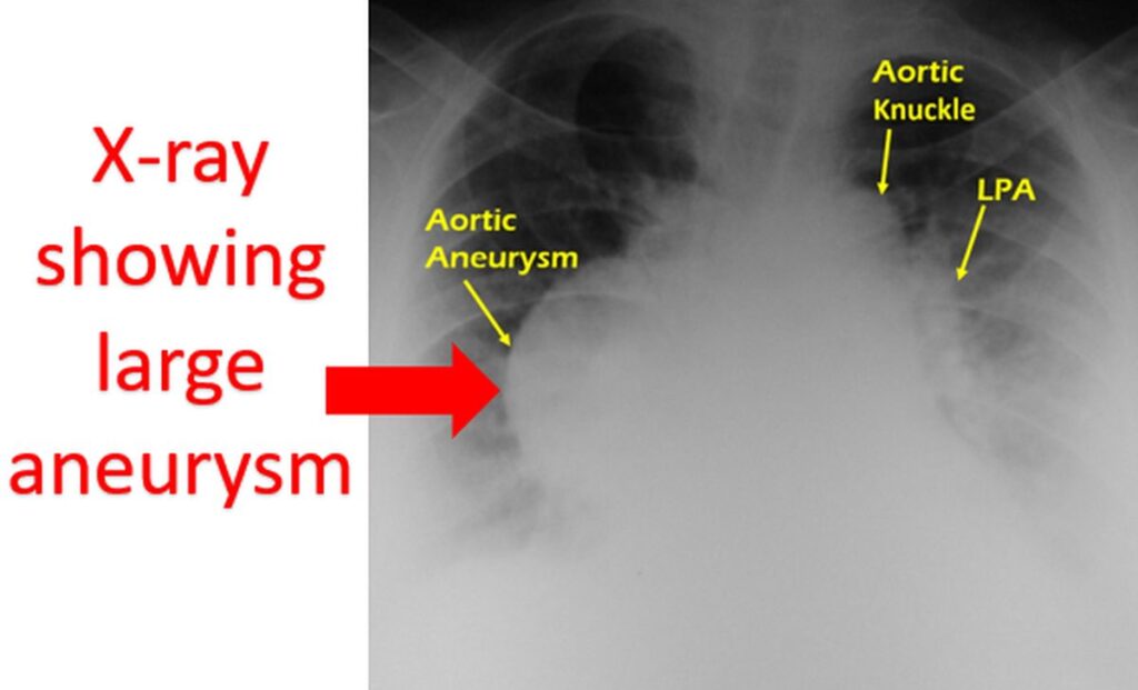
Aneurysm of aorta in the tummy can be imaged very well by an ultrasound scan. Aortic aneurysm in the chest can be noted on X-ray of the chest if it is significantly large. Computed tomographic (CT) scan after injecting contrast medications is the ideal test for aortic aneurysm. Three dimensional reconstruction of CT images give an excellent view of the extent of the aneurysm and location of various branches of the aorta. This is very useful in planning treatment.
Aneurysms can be treated by both surgical repair and by implanting specially made stents inside them to support the vessel wall. Stents can be implanted using a device attached to tubes through small holes made in the blood vessel of the groin. The procedure is done in a cardiac catheterization laboratory, guided by X-ray imaging. Certain aneurysms in the upper chest need a hybrid approach with both surgery and stent placement. This is because blood vessels to the brain arise from the aorta in that region.
Smaller aneurysms need periodic evaluation by imaging tests to see the rate at which they are enlarging. Rapidly enlarging aneurysms need early surgery or stent implantation. Ultrasound scan is a simple test to follow up aneurysm of the aorta in the tummy (AAA). Aneurysms in the chest will need CT scan for follow up. Control of risk factors mentioned initially is very important in reducing the rate of progression.

About The Author
Johnson Francis
Former Professor of Cardiology, Calicut Govt. Medical Kozhikode, Kerala, India. Editor-in-Chief, BMH Medical Journal