What is carbon dioxide angiography?
What is carbon dioxide angiography?
Carbon dioxide angiography is done using carbon dioxide injection into the blood vessel. Angiogram is visualization of a blood vessel by injecting a contrast material into them and imaging them. Usually iodine containing medications known as radio contrast are injected into the blood vessel and continuous X-ray imaging done, to obtain a movie of the blood vessel. Contrast material is needed to show the blood vessel separately from the other body tissue. But usual iodinated radiocontrast medications have a potential risk of affecting kidney function. This is more so in those with already impaired kidney function. It is in this situation that carbon dioxide angiography is usually considered.
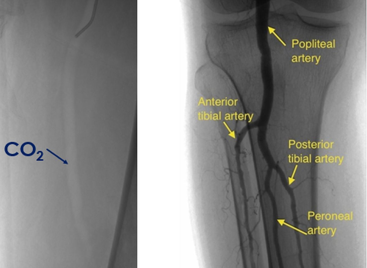
Usual angiograms appear dark while carbon dioxide angiogram looks grey because of the lower density of carbon dioxide compared to tissue. Conventional angiogram is shown beside the CO2 angiogram for comparison of opacification. Please note that the angiograms are not in the same projection. Of course the better contrast with conventional angiogram is obvious. While conventional angiograms look like positive images, carbon dioxide angiograms look like negative images.
Earlier those with kidney problems were considered for magnetic resonance angiography which was also called dyeless angiography. This is because blood vessels can be visualized in MRI with the contrast obtained from moving hydrogen nuclei in water, an important constituent of blood. But for better visualization, gadolinium-based contrasts are often used in magnetic resonance angiography. Gadolinium based contrasts have been associated with a condition called nephrogenic systemic fibrosis in those with kidney failure. Nephrogenic systemic fibrosis is a condition resembling a disease known as scleroderma, with thickening and darkening of large areas of skin. The disease can also involve multiple organs of the body sometimes.
Hence the role of carbon dioxide angiography in those with kidney failure as carbon dioxide is devoid of any such toxicity. As carbon dioxide is highly soluble in blood and rapidly cleared from blood, it is ideally suited as a negative contrast agent in those with kidney failure. But the technique of carbon dioxide angiography is a little more cumbersome than contrast angiography. We need a medical grade carbon dioxide cylinder, three way stop cocks, connector tubings, syringes and a reservoir bag for interim storage of carbon dioxide.
Carbon dioxide angiography is an alternative in those with contrast allergy as carbon dioxide is non-allergenic. Carbon dioxide angiography should not be used for blood vessels of the brain and injections should be below the level of the diaphragm. Diaphragm is the large muscle used for breathing, separating the chest cavity from the tummy. That would mean that carbon dioxide angiography cannot be used in the upper part of aorta or for coronary angiography. Aorta is the large blood vessel arising from the heart. Coronary angiography is visualization of blood vessels of the heart.
Carbon dioxide angiography has also been used to guide angioplasty procedures within the tummy and in the legs. Angioplasty is removal of blocks in blood vessels using balloons attached to thin tubes. Some complications have been reported with carbon dioxide angiography due to formation of vapour lock within the blood vessels. Bleeding at the blood vessel puncture site can occur in carbon dioxide angiography as in conventional angiography. This is prevented by giving good compression after removal of the injecting device. Complications can also be due to contamination with room air which contains nitrogen in plenty and can produce air locks within the blood vessels. Use of a good airtight system and meticulous attention to technique can reduce the complications.
Related Posts


About The Author
Johnson Francis
Former Professor of Cardiology, Calicut Govt. Medical Kozhikode, Kerala, India. Editor-in-Chief, BMH Medical Journal