What is cardiac tamponade?
What is cardiac tamponade?
Cardiac tamponade occurs when fluid collects in the covering of the heart and compresses the heart. Fluid collects between the two layers of the pericardium. Pericardium is the outer covering of the heart with an inner and an outer layer. Fluid collects between these two layers. Fluid can be secondary to infection or bleeding into the pericardial cavity. When fluid collects under tension in the pericardial cavity, usually the right sided chambers collapse and prevent proper filling of the heart.
When the heart is unable to fill properly when it relaxes after a contraction, the amount of blood which can be pumped out in the next contraction also comes down. If it is severe, blood pressure falls and the person may faint. Breathlessness is often associated. Cardiac tamponade is a potentially life threatening condition unless relieved by promptly removing the fluid collection. Fluid can be removed by aspiration with a syringe in case of a dire emergency. When enough time is available, aspiration is done under X-ray imaging or ultrasound guidance.
The cause for the fluid collection should be treated to prevent recurrence. One of the most common causes for cardiac tamponade is bleeding into the pericardial cavity due to a cancerous growth. Primary cancers of the heart are rare and often it is a secondary spread from a nearby organ like lungs or breast. Certain cancers related to blood cells can also cause bleeding into the pericardial cavity and pericardial tamponade. Secondary spread of cancer can occur from a distant organ as well, though less common.
Infections of the pericardium like tuberculosis can also cause cardiac tamponade sometimes. Bacterial infection of heart valves spreading to the pericardium can cause pus in the pericardial cavity. Infections may also be associated with bleeding into the pericardial cavity due to friable cell collections. Certain diseases can cause inflammation of pericardium and fluid collection even without infection. Primary tumours of the pericardium like mesothelioma and lymphoma can also rarely cause cardiac tamponade. In case of lymphoma, there could be involvement of lymph nodes in other regions of the body as well.
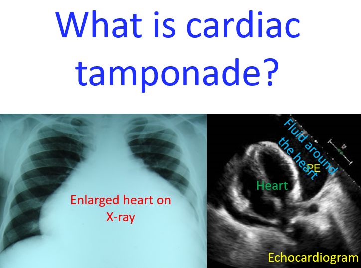
X-ray of chest will show appearance of heart enlargement when there is a lot of fluid collection in the pericardial cavity, known as pericardial effusion. ECG will show a finding called electrical alternans with alternation in the amplitude of waves as the heart swings within the fluid.
Best way to document a large collection of fluid around the heart is ultrasound study known as echocardiography. Now bedside echocardiography with portable echocardiograph is available in the emergency department in all major hospitals.
Bedside ultrasound is very useful in safely removing the fluid collected around the heart without injuring the vital structures as it gives real time images during the procedure. But a more usual way of removing the fluid in major centres when it is not a very dire emergency is to do the aspiration under X-ray imaging guidance in the cardiac catheterization laboratory. This method helps to retain a small flexible tube inside the pericardial cavity for repeated aspiration in case of recollection of fluid. The tube is removed later when the primary disease process is under control.
Related Posts


About The Author
Johnson Francis
Former Professor of Cardiology, Calicut Govt. Medical Kozhikode, Kerala, India. Editor-in-Chief, BMH Medical Journal