How is a 2D echo test done?
How is a 2D echo test done?
2D echo is short form for two dimensional echocardiogram. When echocardiogram, the ultrasound study of the heart, was invented, it was a one dimensional study known as M-Mode echocardiogram. In M-Mode echocardiogram, also known as TM or time motion mode, movement of various structures of the heart was charted along the Y-axis with time elapsed as X-axis. There was also B-mode or brightness mode in which the intensity of echoes from the structures was depicted. 2D echo is a two dimensional representation of echoes received from the structures of the heart. When it is done as live session, it is called real time 2D echo. Further advancement is three dimensional reconstruction, known as 3D echo. Initially it was still 3D reconstructions obtained by post processing. Later when real time 3D was available, some called it 4D imaging as well!
This is an example of an M-Mode echocardiogram of the left ventricle and part of the right ventricle (lower chambers of the heart).
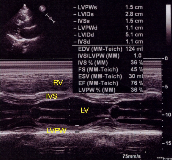
M-Mode echo is mostly used for a quick measurement of left ventricular function in a busy echo lab, though there are many more information which can be gained from it. Most operators are becoming less familiar with M-Mode with the availability of more advanced modes of echo.
In 2D echo, it is almost like viewing the heart with a torch beam in the darkness. Only difference is that instead of the torch beam, you are using an echo probe which sends and receives the ultrasound signals in a sector. Ultrasound is high frequency sound which is not audible to the human ear. The ultrasound beam gets reflected from all the interfaces of the various structures of the heart. Ultrasound has higher velocity in solids than in liquids. So the reflections are mainly from the interfaces of various parts of the heart with the blood in the heart. The reflected waves are collected by the probe and analysed by the computer software in the echo machine to give a real time two dimensional moving image of the heart.
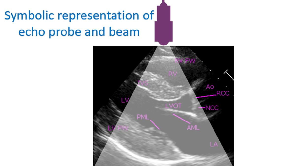
Unlike in M-Mode, it is easier to identify the various structures of the heart in a real time 2D echo so that even an untrained person can get some idea if shown the images. Still good knowledge of the positions of various structures of the heart is needed for correct interpretation. This is more so in complex birth defects of the heart in which sizes and positions of the chambers and valves of the heart may vary much from what is normally expected. Echo beams cannot be sent from all parts of the chest as lungs cover the heart in most regions. Air in the lungs do not allow ultrasound beam to pass into the heart and appears as dense white echoes.
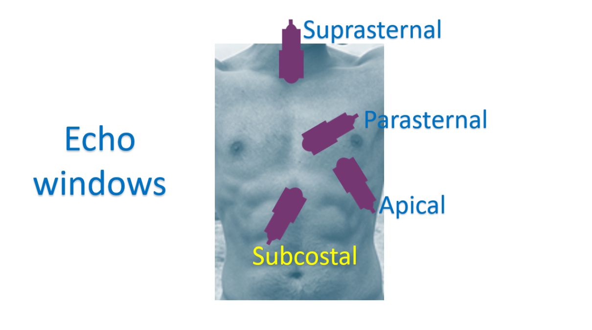
Regions on the chest and upper part of the tummy from which the echo beams can be sent to the heart are known as echo windows. In these regions, lungs usually do not cover the heart much or the heart is very close to the location of the probe. Common sites are the front of the chest near the breast bone, at the place where you feel the heart beats, upper most part of the tummy and in the neck just above the breast bone. These echo windows are called left parasternal, apical, subcostal and suprasternal respectively. Left parasternal means on the left side of the breast bone (sternum). Apical means at the apex of the heart, where the heart beats are felt. Subcostal means just below the rib cage, in the upper end of the tummy (costal means in relation to the ribs). Suprasternal means above the breast bone.
Echo test is usually done with the subject slightly turned to the left side so that the heart becomes closer to the chest wall. Most of the interpretations are done by visualizing the live images, while some may require close scrutiny of frozen images. Both still images and cine loops may be stored in the machine future retrieval. ECG leads may also be connected to get signals for timing along with the echo images. ECG is the electrical recording of the heart. The echo images and videos can be saved to a DVD (digital versatile disc) or pen drive as well, for transfer to another computer. Modern echo machines can also be connected to the hospital information network using LAN (local area network) cables so that physicians can view the echo images and videos on computers in their office as well. A picture archiving and communication system (PACS) can also be used in the hospital for storing and retrieval of echo information.
Related Posts


About The Author
Johnson Francis
Former Professor of Cardiology, Calicut Govt. Medical Kozhikode, Kerala, India. Editor-in-Chief, BMH Medical Journal