How is an ECG taken?
How is an ECG taken?
ECG is short for electrocardiogram, the recording of the electrical activity of the heart. It is recorded by placing metallic contacts known as electrodes in specified parts of the limbs and chest and connecting them to the ECG machine known as an electrocardiograph. The electrical signals from the heart detected on the surface of the body are tiny, in the range of millivolts. The ECG machine amplifies these tiny signals and records the variation in the signals with time on a moving graph paper. This record of the electrical activity of the heart recorded from the body surface is the ECG.
As these signals are tiny, it is essential to avoid interferences from other electrical signals in the vicinity and from the body itself. An electrical interference which can arise from within the body is the electrical activity of contracting muscles. So it is essential for the person to stay relaxed during ECG recording. Movements of limbs or even tremor due to undue anxiety can produce a lot unwanted electrical signals on the ECG, sometimes making interpretation difficult. This is quite common in the case of small children who are often upset by the multiple wires being fixed on the body. Hence recording ECG in children is done either during natural sleep or after sedation with suitable medication. Electrical equipment in the vicinity which can generate electrical interference may have be switched off during the recording, especially in places like the hospital intensive care unit.
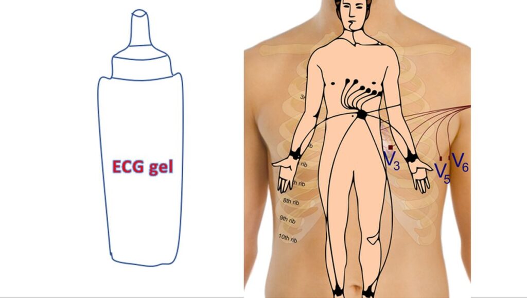
To ensure good electrical contact between the recording electrode and the skin, an electrode gel which is a good conductor of electricity will be applied at locations of skin contact.
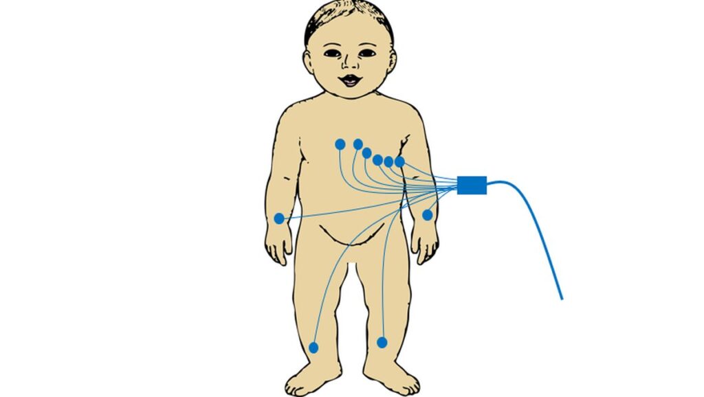
If the designated region is too hair, it may be necessary to shave the region to get a good electrical contact. In young children, small sticky electrodes may be used instead of the electrode clips and suction electrodes for the chest. Applying a suction electrode on the chest of a small infant can produce mild bruising of skin. Moreover there will not be enough space on the chest for applying multiple adult sized suction electrodes.
Usually four electrode clips are placed on the four extremities, at the wrists and ankles. Six suction cup electrodes are placed over specified locations of the chest. These electrodes are connected to the machine through an ECG cable. Within the machine, different combinations of electrodes can be chosen to record different ‘ECG leads’. In standard 12 lead ECG, 6 limb leads and 6 chest leads are recorded. Sometimes additional electrodes may be placed on the chest, known as right chest leads, when specific abnormalities are suspected or in the rare situation of the heart being on the right side.
ECG is recorded on a heat sensitive thermal coated paper. The tracings have a tendency to fade as time passes. So it is advisable to have a photocopy of the tracing for long term record. Many take snapshots on their mobile phones these days to have a virtual copy! Some types of ECG equipment like certain treadmill exercise ECG recorders can print on usual type of paper which does not fade with time. But most other treadmill exercise ECG recordings also fade with time and needs photocopy for long term storage.
Related Posts


About The Author
Johnson Francis
Former Professor of Cardiology, Calicut Govt. Medical Kozhikode, Kerala, India. Editor-in-Chief, BMH Medical Journal