How is device closure of ASD done?
How is device closure of ASD done?
ASD is short form for atrial septal defect. Atrial septal defect is a birth defect of the heart. It is a hole in the wall between the two upper chambers of the heart. This causes entry of blood from the left atrium (left upper chamber) to the right atrium (right upper chamber). When the flow is large, it can increase the pressure in the lungs and cause damage to blood vessels of the lungs in the long run. Though small ASDs can be left alone, large ASDs used to be closed by open heart surgeries earlier. Now some types of ASDs can be closed without surgery, using a device introduced through the blood vessel of the groin. This procedure is called a device closure of ASD.
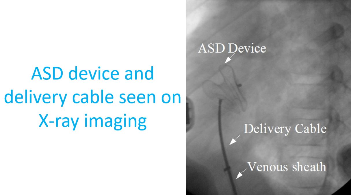
All ASDs are not suitable for device closure. The ASD device is folded like an umbrella and introduced into the heart using a tube known as catheter. ASD device has two discs like a clam shell, one for the left side of the defect and another for the right side, with a small connection link in between. The folded device loaded inside a tube is inserted through a small hole made in the skin of the groin under local anaesthesia. It is guided to the heart using live X-ray imaging in a special facility room called cardiac catheterization laboratory.
Once the device has crossed the ASD, position is further confirmed by echocardiogram, ultrasound imaging of the heart. After confirmation of position, first the left sided disc is opened from the catheter used for introduction. Later the right sided disc is also opened. Once both discs are in position, echocardiogram is done again to check for any remaining leaks and whether the position of the device is acceptable. It should not be interfering with the function of the nearby valves. If everything is fine, the device is released from the delivery system. Then the delivery system is withdrawn and taken out.
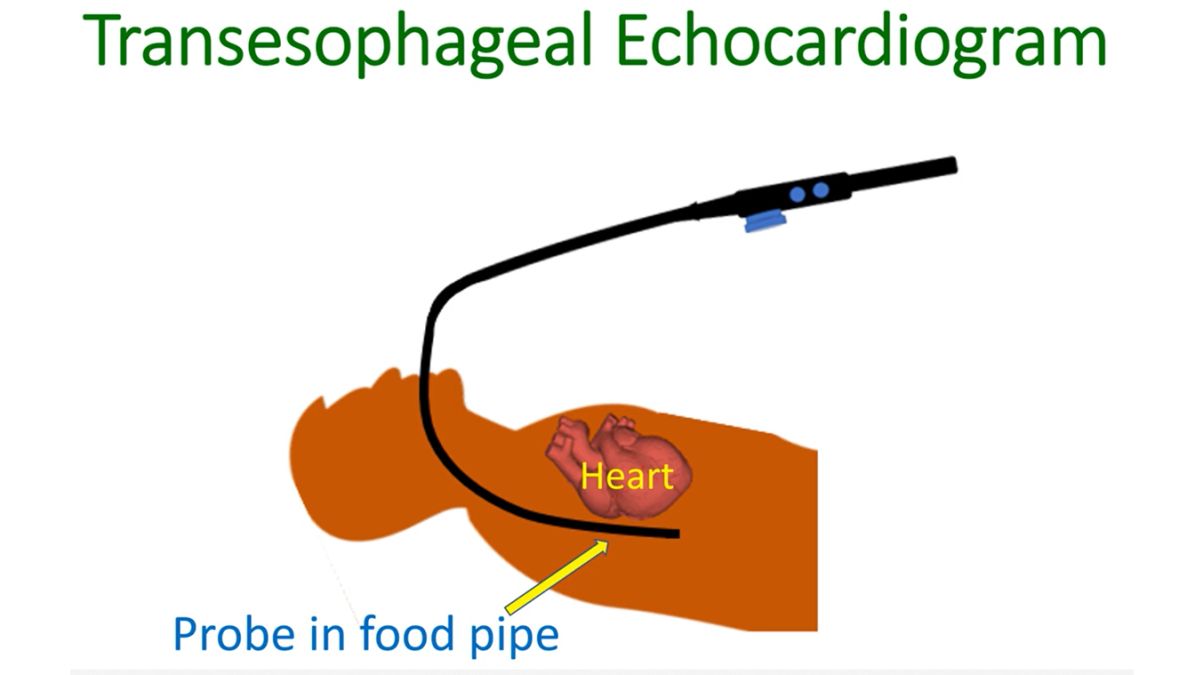
The suitability of ASD for device closure is decided by an echocardiogram test. In addition to a routine echocardiogram done from the chest, a special echocardiogram known as transesophageal echocardiogram (TEE) is used to assess the finer details of the ASD. TEE is done using a probe introduced into the food pipe, just behind the heart. The exact location of the defect, distance from other important parts of the heart like heart valves and aorta are measured. Aorta is the largest blood vessel in the body carrying oxygenated blood. A reasonable distance from all these vital structures is needed to place the device without immediate and long term complications.
While loading the folded device into the delivery system, special care is taken to avoid introduction of air bubbles along with it. This is done by loading the device under water in a tray. The delivery system is flushed well before introduction into the blood vessel to remove any air bubbles. If air enters the blood vessels, it can go and block some blood vessel in the brain, heart or other vital organs.
Device closure of ASD can be done under local anaesthesia in adults. In children who are afraid of the procedure, general anaesthesia may be used. TEE may be used during the procedure to monitor the exact position of the device in the heart to ensure proper closure of the defect. TEE during procedure will also tell if the device is impinging on any nearby important heart structures. If any such problem is found, the device can be repositioned or removed if necessary. If the procedure is successful, it avoids a major open heart surgery.
Related Posts


About The Author
Johnson Francis
Former Professor of Cardiology, Calicut Govt. Medical Kozhikode, Kerala, India. Editor-in-Chief, BMH Medical Journal