What is aortic regurgitation?
What is aortic regurgitation?
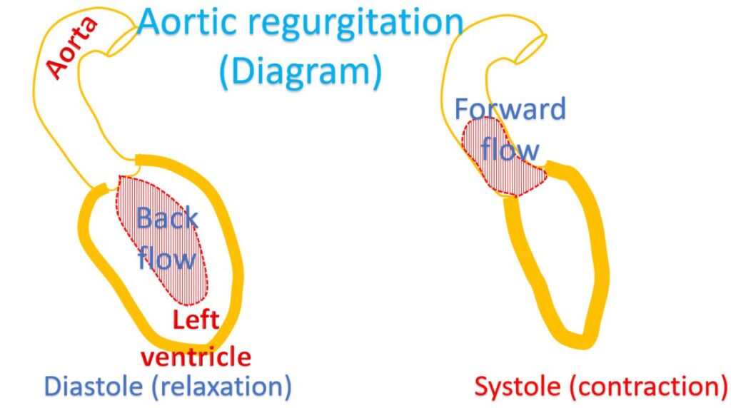
Aortic regurgitation is leakage in the aortic valve which separates aorta from the left ventricle. Aorta is the largest blood vessel into which left ventricle, the left sided muscular chamber of the heart pumps blood. The normal function of the aortic valve is to prevent back flow of blood from the aorta to the left ventricle. When it is leaky, it leads to volume overload of the left ventricle, which has to pump more blood in its next contraction. Part of it returns back to the left ventricle when it relaxes and the cycle continues.
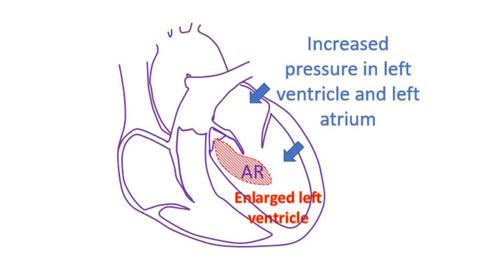
The overload to the left ventricle leads to its enlargement and gradual increase in the wall thickness to take care of the extra work load. The pressures in the left ventricle also rises due to the extra blood volume. When the leak is severe the left ventricle fails and the back pressure is transmitted to the left atrium, the upper chamber which receives oxygenated blood from the lungs. The raised pressure in the left atrium is in turn transmitted to the blood vessels of the lungs. When the pressure in the blood vessels of the lungs go beyond a certain limit, usually 25 mm Hg, fluid collects in the air cavities of the lungs. This is known as pulmonary edema. The lungs become heavy and oxygen exchange is defective. Breathlessness occurs, initially during exercise and sometimes in sleep. This situation is known as heart failure.
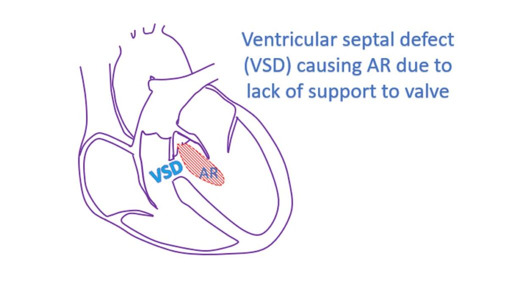
Aortic regurgitation can occur in a disease known as rheumatic fever, which can affect other valves as well. A birth defect of the aortic valve in which the valve has only two cusps instead of three (bicuspid aortic valve) can also lead to aortic regurgitation. Infections of the aortic valve damage the valve leaflets leading to perforations and severe aortic regurgitation. Degenerative changes as age advances can also damage the aortic valve and cause regurgitation. Other birth defects like a ventricular septal defect (hole in the wall between the lower chambers of the heart) can also be associated with aortic regurgitation. This occurs because the support to the aortic valve leaflets from below is lost due to the defect just below it.
Undue awareness of heart beats known as palpitation can also occur in aortic regurgitation in addition to breathlessness. This is because of the enlarged volume overloaded left ventricle which beats more forcefully. Some persons may experience chest pain, especially while in bed at night in aortic regurgitation.
Aortic regurgitation can be detected using a stethoscope as there is a specific heart murmur which occurs when the heart relaxes after contraction, at which time the leak occur occurs. Important tests done in case of aortic regurgitation are ECG, X-ray of the chest and echocardiogram or ultrasound study of the heart. ECG and X-ray will give indications of enlargement of the left ventricle. Echocardiogram will show the leak and the severity can be quantified. It will also give an estimate of the sizes of the heart chambers and the pumping function of the left ventricle. Specific findings may be noted in case of infections of the aortic valve.
If there is infection of the aortic valve, blood culture is obtained to identify the micro organism responsible and to decide on appropriate treatment. A prolonged course of anti microbial treatment is needed for infections of the aortic valve. Occasionally if the valve is severely damaged or when there is a complication, the valve may have to be replaced by surgery to eradicate the infection.
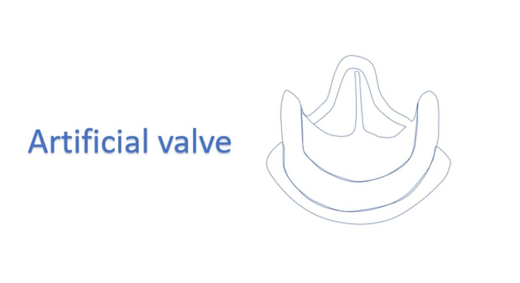
Mild and moderate aortic regurgitation is usually asymptomatic and detected as an incidental finding on a medical evaluation for some other reason. Severe aortic regurgitation can also be asymptomatic if it has progressed gradually. Those with severe aortic regurgitation can be given medications which enlarge the peripheral blood vessels so that more blood flows forward into them than back into the left ventricle. This will reduce the load of the left ventricle. When there is no response to medical management aortic valve can be replaced by surgery. Both mechanical valve and bioprosthetic valves are available for this purpose. If a mechanical valve is placed, the person will need lifelong medications to prevent clotting of the artificial valve. Bioprosthetic valves also need medications to prevent clot formation for the initial few months. They have much lesser tendency for clot formation as they are made from processed animal valves.
Related Posts


About The Author
Johnson Francis
Former Professor of Cardiology, Calicut Govt. Medical Kozhikode, Kerala, India. Editor-in-Chief, BMH Medical Journal