What is Blalock – Taussig (BT shunt) operation?
What is Blalock – Taussig (BT shunt) operation?
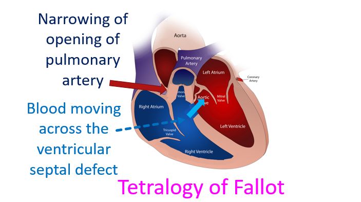
Blalock – Taussig shunt is an operation done to improve the oxygen levels in blood of children with certain birth defects of the heart. Typical example is a condition known as tetralogy of Fallot with four defects. Components of tetralogy of Fallot are a defect in the wall between the lower chambers known as ventricular septal defect, narrowing of the opening to the pulmonary artery known as pulmonary stenosis, thickening of the wall of the right ventricle and overriding of the ventricular septal defect by the aorta.
Right ventricle is the right lower chamber of the heart which pumps blood to the lungs for oxygenation. Pulmonary artery is the blood vessel which takes blood to the lungs from the right ventricle. Aorta is the large blood vessel which carries oxygenated blood to the whole body from the left ventricle. Left ventricle is the lower left chamber of the heart.
In tetralogy of Fallot and similar birth defects of the heart, blood flow to lungs is reduced due to narrowing of the opening to the pulmonary artery. Oxygen poor blood in the right ventricle moves across the ventricular septal defect to the aorta. This leads to oxygen poor blood reaching the whole body, producing a bluish colour known as cyanosis. Hence this group of diseases are called cyanotic congenital heart diseases.
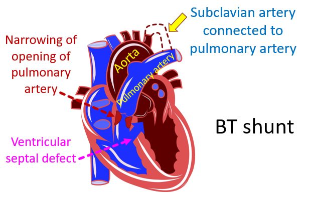
Blalock – Taussig shunt known in short as BT shunt creates a connection between a branch of the aorta and a branch of the pulmonary artery. Blood in the aorta at high pressure flows into the pulmonary artery at lower pressure. This increases the blood flow to the lungs and improves oxygen content of the blood reaching the left atrium the left upper chamber. In turn blood from the left atrium reaches the left ventricle and is pumped into the aorta. Hence BT shunt increases the oxygen content of the blood reaching the body and decreases cyanosis.
BT shunt is a temporary procedure for the small baby not fit for a major operation to totally correct the combination of birth defects in tetralogy of Fallot. When the baby is better, total correction is done by an open heart surgery. In selected cases, total correction can be done as the first procedure without going for a BT shunt, in advanced centres. Hence the number of BT shunts for tetralogy of Fallot has come down. Moreover there are also other procedures which can be done using tubes passed through blood vessels, which can produce the similar results. BT shunt may still be used for more complex birth defects which cannot be repaired fully in early childhood.
In BT shunt, because the aorta at high pressure is connected to the pulmonary artery at low pressure, sometimes blood flow to the lungs can be much more than is needed. This can damage blood vessels of the lungs in the long run. Hence modifications of the BT shunt known as modified BT shunt has been developed. In original BT shunt, a branch of the aorta known as subclavian artery, which takes blood to the arm is used to take blood to the pulmonary artery.
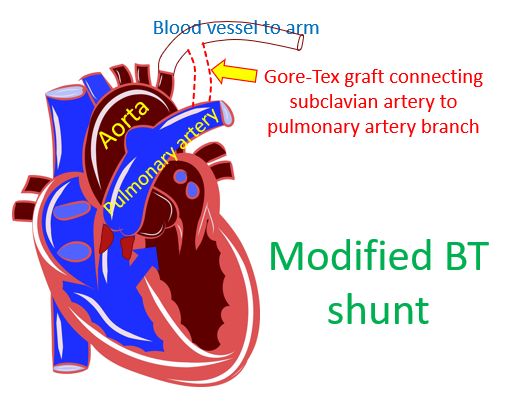
This can reduce the blood supply to the arm. In modified BT shunt an extra tube known as a Gore-Tex graft is used to connect the subclavian artery to the pulmonary artery. This takes only a part of the blood going to the arm and hence the blood pressure in the lungs does not go very high. Moreover, the blood flow to the arm is also preserved to some extent. Classical BT shunt has been abandoned and only modified BT shunts are being done now.
The modified BT shunt can be removed at the time of complete repair of tetralogy of Fallot. This prevents future problems due to potential decrease in blood supply to the arm and increased blood supply to the lungs. There is also a small risk of infection if the shunt remains there lifelong. Alternatively, the modified BT shunt can be occluded by a coil or device introduced through blood vessels under the guidance of X-ray imaging in a cardiac catheterization laboratory.
Related Posts


About The Author
Johnson Francis
Former Professor of Cardiology, Calicut Govt. Medical Kozhikode, Kerala, India. Editor-in-Chief, BMH Medical Journal