What is Ebstein’s anomaly?
What is Ebstein’s anomaly?
Ebstein’s anomaly is a birth defect of the tricuspid valve, which may manifest soon after birth or more commonly later in life. Tricuspid valve is the valve between the right atrium and right ventricle. Right atrium is the right upper chamber and right ventricle the right lower chamber of the heart. In Ebstein’s anomaly, two of the three leaflets of tricuspid valve are displaced further into the right ventricle. So a part of the right ventricle becomes part of the right atrium in effect.
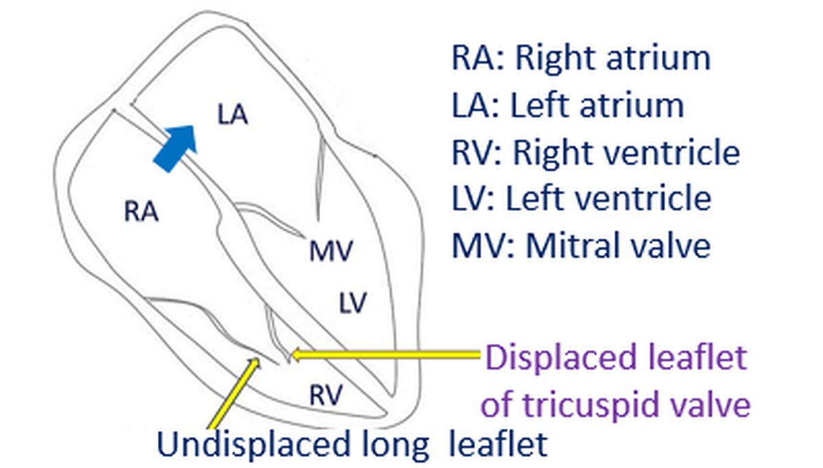
The tricuspid valve in Ebstein’s anomaly can have severe leak or obstruction, both of which leads to enlargement of the right atrium. As part of the right ventricle is taken away by the right atrium, right ventricle is small in Ebstein’s anomaly. Sometimes there is a defect in the wall between the two upper chambers, known as atrial septal defect. As the pressure in the right atrium is usually higher than that in the left atrium in Ebstein’s anomaly, blood shunts from right atrium to left atrium.
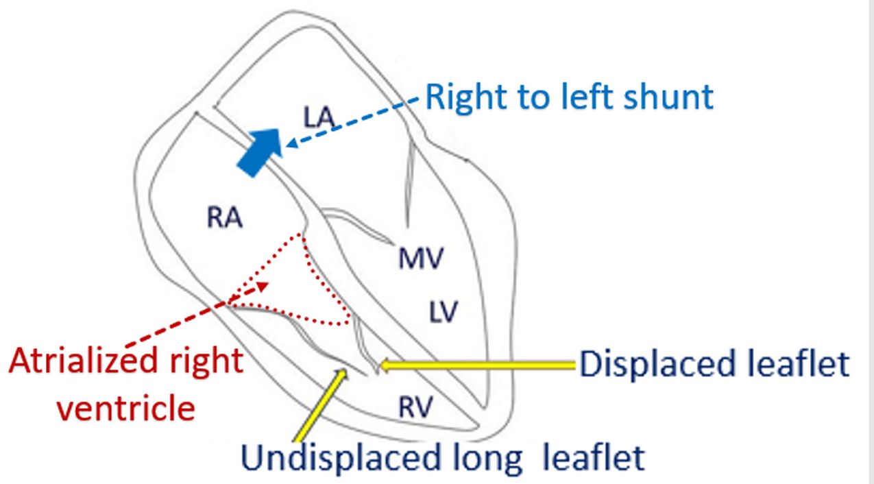
Right atrium receives blood from the great veins of the body which return oxygen poor blood for oxygenation from the lungs. When right atrial blood mixes with left atrial blood, the oxygen content of left atrial blood decreases. This blood flows into the left ventricle, the lower chamber and is pumped out into the aorta. Aorta is the largest blood vessel taking blood to the whole body. Oxygen content of blood is less when there is a mixing with right atrial blood and hence bluish discoloration of skin, lips and tongue may occur.
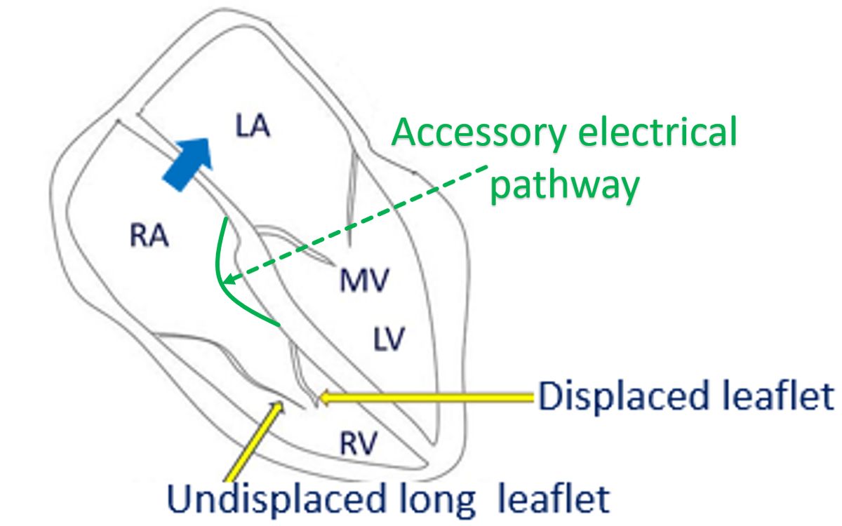
Enlarged right atrium and the part of the right ventricle included in the right atrium can be the focus of abnormal heart rhythms, which could be fast and dangerous. In addition, there could be an accessory electrical conduction pathway from the right atrium to right ventricle. The accessory pathway also predisposes to fast heart rhythm abnormalities. Some of the abnormal rhythms originating from the lower chambers could cause sudden death rarely.
Mild varieties with Ebstein’s anomaly may not produce many symptoms and present in adult life, often detected during routine medical examination for another reason. Severe varieties can manifest in infancy with bluish discoloration of the skin, lips and tongue. Severe varieties may be treated with an open heart surgery which involves a special repair of the abnormal tricuspid valve known as cone procedure. But surgery may not decrease the chance for heart rhythm abnormalities. In fact new focus of heart rhythm abnormalities may arise in the surgical scar.
The abnormality in the electrical conduction will be manifest in the ECG, the recording of the electrical activity of the heart from the body surface. Those with recurrent heart rhythm abnormalities will need a special test known as electrophysiology study (EP study). In electrophysiology study, small electrode wires are introduced into the heart through the blood vessels of groin and neck. Recording of the electrical activity from within the heart helps in localizing the accessory conduction pathways.
Once the accessory pathways are localized, they can be inactivated by tiny burns produced by high frequency current delivered to the precise location. This procedure is known as radiofrequency catheter ablation (RF ablation). RF ablation abolishes conduction in the accessory pathway and thus cures the heart rhythm abnormality. EP is study is done in all cases taken for surgery in some centres.
Related Posts


About The Author
Johnson Francis
Former Professor of Cardiology, Calicut Govt. Medical Kozhikode, Kerala, India. Editor-in-Chief, BMH Medical Journal