What is ECG imaging (ECGi)?
What is ECG imaging (ECGi)?
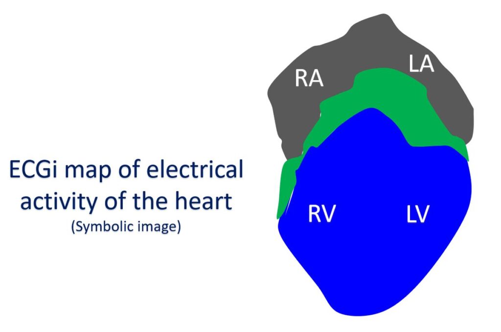
ECG is the recording of the electrical activity of the heart from the body surface. ECG imaging (ECGi) uses a special vest with multiple electrical contacts over the chest to record a three dimensional map of the electrical activity of the heart. Previously electrical mapping of the heart was done using small wires introduced into the heart. ECGi uses a 252 electrode vest to record a similar map non-invasively. CardioInsight™ uses a single use, disposable, multi-electrode mapping vest to acquire electrophysiological data from the body surface. It combines the surface ECG signals with computed tomography (CT) scan data to produce and display simultaneous 3-D cardiac maps from all four chambers of the heart.
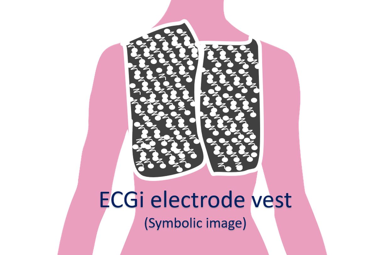
The vest has three multi-electrode panels for front left, front right and back. Connectors from the electrode vest is connected to the CardioInsight workstation. The chest is shaved and prepared with a special solution. The front left panel is applied first followed by the front right and back panels. Panels are secured with medical tape. CT scan is done with vest on. Signals from the 252 electrodes are obtained and integrated with the CT data. Alternatively, the ECG data can be acquired first and CT images obtained shortly after.
The workstation provides various types of high resolution maps of the electrical activity of the heart which can be interpreted by the heart rhythm specialist. ECGi uses a standardized workflow and it has been suggested that signals should be checked manually to avoid automatic processing errors. ECGi helps us to understand the mechanism of heart rhythm disorders and guide treatment procedures.
ECGi has been used to find the site of origin of heart rhythm disorders originating from the lower chambers of the heart known as premature ventricular complexes and ventricular tachycardia. Another novel application was identification of the site of origin by ECGi followed by treatment with external beam radiation used in cancer treatment. This avoids entering the heart with wires as is usually done for a procedure known as radiofrequency catheter ablation. But as of now, the accuracy is not that high so that it is advised only in persons at very high risk for conventional catheter ablation as it cannot give highly precise images and treatment delivery.
Another condition in which ECGi has been used is an abnormal rhythm originating from the upper chambers of the heart known as atrial fibrillation. Atrial fibrillation is a fast irregular rhythm which is the commonest sustained heart rhythm abnormality. Chances of atrial fibrillation increases as age advances. ECGi helps in identifying the type of electrical activity in the upper chambers of the heart so that regions of interest can be selected for catheter ablation.
Related Posts


About The Author
Johnson Francis
Former Professor of Cardiology, Calicut Govt. Medical Kozhikode, Kerala, India. Editor-in-Chief, BMH Medical Journal