What is mitral valve prolapse (MVP)?
What is mitral valve prolapse (MVP)?
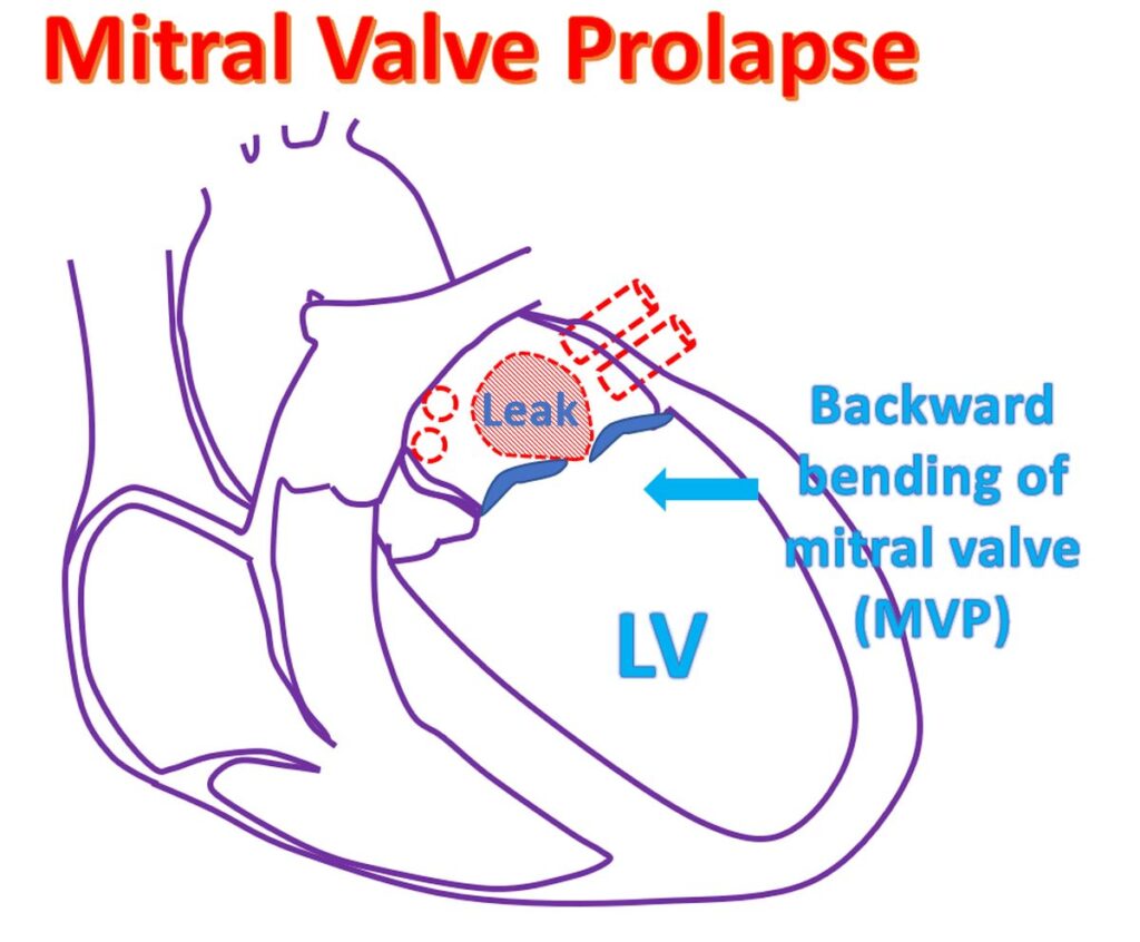
Mitral valve prolapse (MVP) is bending backwards of the mitral valve as it closes when the left ventricle contracts. Left ventricle is the lower left chamber which pumps oxygenated blood to the whole body. Mitral valve is the valve between the left upper and lower chambers of the heart. Too much of bending may produce a leak in the mitral valve. When the mitral valve is leaky, blood flows back into the left atrium, when the left ventricle contracts. Left atrium is the upper chamber from which the left ventricle receives blood when it relaxes after a contraction.
Left atrium in turn receives oxygenated blood from the lungs through the pulmonary veins. When the leak is severe, the pressure in the left atrium rises. Left ventricle enlarges to take care of the extra work load as the leaking blood returns back to the left ventricle when it relaxes after the contraction. Increase in pressure in the left atrium is transmitted back to the pulmonary veins and hence to the lungs. Leak in the mitral valve is called mitral regurgitation.
Increase in pressure in the pulmonary veins leads to collection of fluid in the tiny air spaces of the lungs known as alveoli. This is known as pulmonary edema. Fluid in the alveoli makes oxygenation of blood difficult and the person becomes breathless. This occurs only in the late stages as the left atrium and left ventricle can enlarge and take the extra load initially without increasing the pressure within.
Mitral valve prolapse is often seen in very lean individuals and can be associated with abnormalities of the skeletal system like sideways or forward bends of spine (kyphoscoliosis). Many of these individuals have a lot of associated anxiety and palpitation which may be disproportionate to the severity of leak in the valve. This has been called mitral valve prolapse syndrome. Mitral valve is often thickened and redundant due to extra length in persons with mitral valve prolapse.
Usual tests done for a suspected case of mitral valve prolapse are ECG, X-ray of the chest and echocardiogram (ultrasound study of the heart). ECG will tell if there is an abnormal rhythm of the heart. It will also give an indication about enlargement of the heart chambers. Echocardiogram will show the prolapse of the mitral valve and the amount of leak. Increase in size of heart chambers can also be noted. Pumping function of the left ventricle can be estimated by an echocardiogram.
Mild cases need only pacification for relief of anxiety and occasionally medications to relieve anxiety and palpitation (undue awareness of heart beats). If there is a gross abnormality in heart rhythm, it may require medications. Those in heart failure due to severe leak needs medications for heart failure. Occasionally the abnormal mitral valve may get infected and require prolonged treatment with antibiotics.
A valve with severe leak producing breathlessness and heart failure can be repaired or replaced. These are open heart surgeries using heart lung machine and require opening up of the chest. Minimally invasive surgery with small skin openings are also available in selected situations. Valve replacement can be done with either a mechanical valve or biological bioprosthetic valves. Mechanical valves will require lifelong medications to prevent the formation of clots within them. Regular blood tests to monitor clotting function are needed while on medications which prevent clotting.
A technique for implanting a clip between the mitral valve leaflets with a device introduced through a small hole made in the groin skin is a new option for treating severe leaks in the mitral valve. The device is guided into the heart using X-ray imaging equipment and is done in a special procedure room known as cardiac catheterization laboratory (cathlab). This procedure does not require opening up of the chest.
Related Posts


About The Author
Johnson Francis
Former Professor of Cardiology, Calicut Govt. Medical Kozhikode, Kerala, India. Editor-in-Chief, BMH Medical Journal