What is myocarditis?
What is myocarditis?
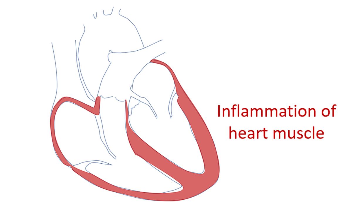
Myocarditis is the inflammation of the heart muscle. It can be caused by various causes like infections with viruses, bacteria, fungi and parasites. Myocarditis can sometimes occur as an allergic reaction to certain medications and vaccines. Certain disorders of the immune system like systemic lupus erythematosus (SLE) may also cause myocarditis. Radiation given for the treatment of cancer in nearby organs like breast and lungs can cause myocarditis rarely. This is rare now-a-days because of advanced technology which can deliver radiation without much collateral damage.
Symptoms of myocarditis may be similar to a heart attack with chest pain, breathlessness and rapid irregular heart beats. But onset of symptoms is usually not so abrupt as in a heart attack. Symptoms of the underlying infection like fever, generalized body aches and head ache may also be noted along with specific symptoms of myocarditis. In advanced cases with heart failure, fluid can collect under the skin, especially in the legs.
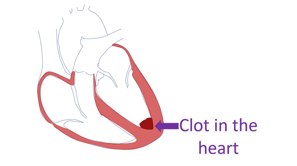
Myocarditis damages the heart muscle, which can be temporary or permanent. Damage to the heart muscle leads to defective pumping function of the heart and heart failure. Heart muscle damage can also initiate abnormal heart rhythms (cardiac arrhythmia) which can sometimes be quite dangerous. Very serious abnormalities of the heart rhythm can rarely lead on to cardiac arrest. Blood clots can form inside the heart in the region of the damaged heart muscle. These clots can move out in the circulation and block blood vessels elsewhere. If it blocks a blood vessel of the brain, a stroke with weakness of one side of the body may occur. Clots can also enter the blood vessels of the heart itself and block them, leading to a heart attack in addition to myocarditis.
Usual tests done in case of suspected myocarditis are similar to those done for other heart diseases. An ECG (recording of the electrical activity of the heart) is the test done most often. It will show changes if there is damage to the heart muscle and abnormalities in heart rhythm. A supportive test when there is damage to heart muscle is a blood test known as cardiac troponin. Cardiac troponins are elevated when there is heart muscle damage due to any cause.
An X-ray of chest will show if the heart chambers have enlarged due to weakening by myocarditis. If the person has gone into heart failure, it will show collection of fluid within the small airspaces of the lungs. This condition is known as pulmonary edema and interferes with oxygen exchange in the lungs. Breathlessness and fall in blood oxygen levels are associated with this finding on X-ray of the chest.
Echocardiogram (ultrasound study of the heart) is another widely used test for the evaluation of heart function. It can be done at the bedside in the emergency room as well as in the ward or intensive care unit. It gives a rapid assessment of the function of heart muscles, function of heart valves and presence of clots within the heart if any. Repeat studies can be done during treatment to assess the progress or deterioration in heart function. Any collection of fluid around the heart can also be identified and quantified.
Other than cardiac troponin blood test, other blood tests are needed to look at the underlying cause for the myocarditis. Antibodies to viruses and bacteria are found in corresponding infections. When the immune system is abnormal there are other antibodies which can be found in the blood. General blood tests to assess the body status are also done as in any other infections.
Magnetic resonance imaging of the heart (Cardiac MRI) is another important test in the setting of myocarditis. It can quantify the damage to the heart muscle and pumping function much better than the echocardiogram. But it is often difficult to obtain in a sick person and cardiac MRI software may not be there in all MRI machines, leave alone the technical skills for interpretation. Still cardiac MRI is an important test in suspected myocarditis as it can indicate the presence of tiny amounts of fluid between the heart muscles cells (myocardial edema).
In some cases, especially in advanced centres a test known as cardiac catherization and heart muscle biopsy may be done to confirm myocarditis. Cardiac catheterization involves the introduction of small tubes through tiny holes made in the skin under local anaesthesia. These tubes known as catheters are guided to the heart using X-ray imaging equipment in a special room known as cardiac catheterization laboratory (cathlab). Tiny amounts of heart muscle can be taken out with specialized devices for study under the microscope (biopsy).
Treatment of myocarditis involves treatment of the causative infection as well as supportive treatment of the heart muscle damage. If heart failure is present, usual medications for heart failure are given. Heart rhythm abnormalities are treated with medications. Medications to suppress the immune system may be added if there is a fault in the immune system causing heart muscle damage. Advanced treatment options like ventricular assist devices to support the damaged heart muscle or extracorporeal membrane oxygenator (ECMO) to support the function of heart and lungs, may be need in some cases.
Many cases of myocarditis recover fully and they return to normal life, though medical follow up may be needed. A few persons may have persistent damage to the heart muscle weakening its pumping function. These persons will need long term medications in addition to regular medical follow up. A small number with persistent severe heart muscle damage not responding to medications may also be considered for heart transplantation.
Related Posts


About The Author
Johnson Francis
Former Professor of Cardiology, Calicut Govt. Medical Kozhikode, Kerala, India. Editor-in-Chief, BMH Medical Journal