What is pulmonary stenosis?
What is pulmonary stenosis?
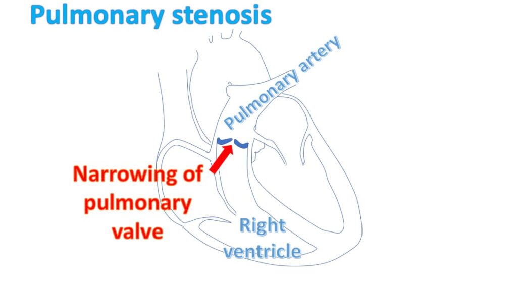
Pulmonary stenosis is narrowing of the valve between the right ventricle and pulmonary artery. Pulmonary artery is the large blood vessel carrying blood to the lungs for oxygen enrichment. Right ventricle is the right lower chamber of the heart which pumps blood to the lungs. Pulmonary valve prevents backflow of blood from the pulmonary artery into the right ventricle when it relaxes after a contraction.
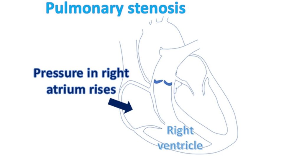
When the pulmonary valve is narrowed, right ventricle finds it increasingly difficult to pump blood into the pulmonary artery. The right ventricular muscle becomes gradually thickened (right ventricular hypertrophy) in an attempt overcome the obstruction. But when the pulmonary stenosis is very severe, right ventricle is unable to overcome the obstruction and gradually fails due the overwork. When right ventricle fails, the back pressure is transmitted to the right atrium, the right upper chamber of the heart.
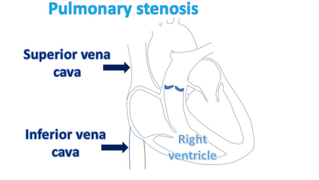
Right atrium enlarges due to the higher pressure inside. This in turn is transmitted to the great veins known as superior and inferior vena cava. Superior vena cava drains the blood from head and neck to the right atrium, to be taken to lungs for oxygen enrichment. Inferior vena cava drains blood from the lower part of the body for transport to the lungs.
Congestion of the superior vena cava manifests as dilated veins in the neck. Congestion of the inferior vena cava causes enlargement of the liver, and collection of fluid under the skin of the legs (edema). In late stages fluid also collects inside the tummy. This state is known as right heart failure. Liver and kidney function may also deteriorate in advanced cases due to the back pressure of blood.
Usual tests done in case of pulmonary stenosis are ECG, X-ray of the chest and echocardiogram (ultrasound study of the heart). Echocardiogram will show thickening and narrowing of the pulmonary valve. The pressure gradient across the valve can be estimated by Doppler echocardiography. This will give an estimate of the severity of obstruction. Right ventricular and right atrial status can also be documented by echocardiography. Pulmonary stenosis is usually caused by a birth defect.
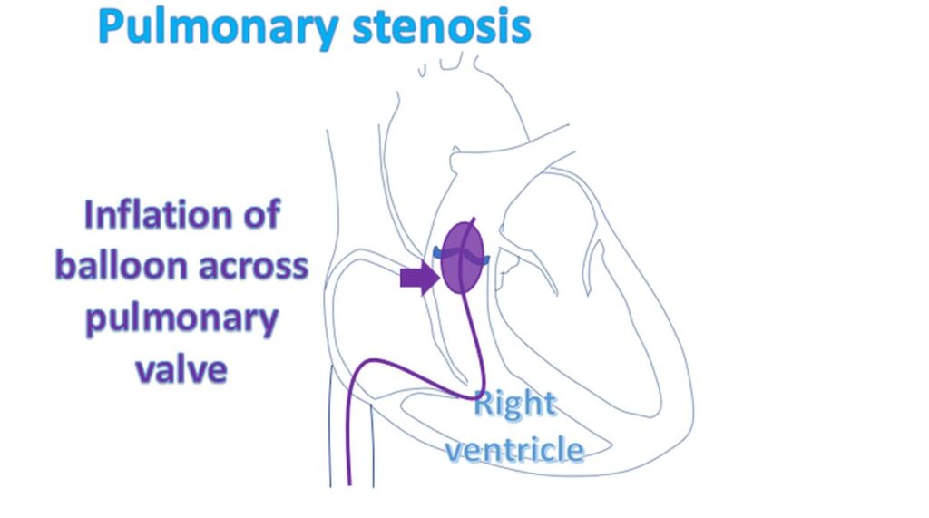
In severe cases, an obstructed pulmonary valve can be opened up by a procedure known as balloon pulmonary valvotomy. In this procedure, a balloon attached to a tube known as balloon catheter, is introduced through a small hole in the groin skin. The balloon is guided across the pulmonary valve using X-ray imaging. When it is across the valve, the balloon is inflated, relieving the obstruction. The procedure is done under local anaesthesia and there is no need to open up the chest.
In the past, surgical pulmonary valvotomy was done by opening the chest under anaesthesia. It is seldom done now a days, unless there are other associated birth defects which need repair by opening the heart.
Related Posts


About The Author
Johnson Francis
Former Professor of Cardiology, Calicut Govt. Medical Kozhikode, Kerala, India. Editor-in-Chief, BMH Medical Journal