What is RF ablation?
What is RF ablation?
RF ablation is short form for radiofrequency ablation. A more complete term with respect to the treatment of heart rhythm disorders is radiofrequency catheter ablation. RF ablation uses very high frequency electrical energy in the range used for radio transmission. While the energy is radiated as radio waves in radio transmission, in RF ablation, it is delivered to small regions of the heart using specialized wires known as ablation catheters. This produces a tiny superficial burn in the region which abolishes the abnormal electrical activity in the heart.
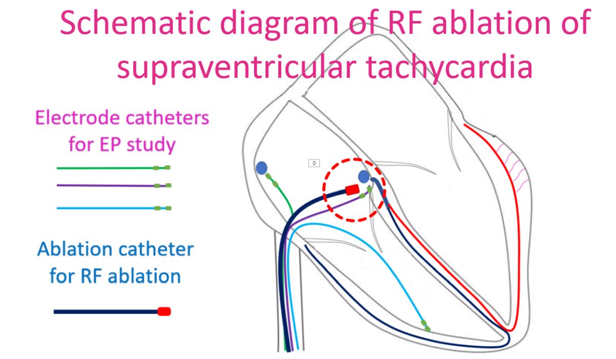
An electrophysiology study (EP study) is done prior to RF ablation to identify the source and mechanism of the abnormal electrical activity or conduction within the heart which causes the heart rhythm abnormality. EP study uses multi-electrode wires introduced into the heart chambers under local anaesthesia, through the blood vessels of the neck and groin. A repeat electrophysiology study is usually done after the RF ablation to make sure that there is no likelihood of recurrence of the heart rhythm abnormality.
Commonest type of heart rhythm abnormality for which RF ablation is done is a fast regular rhythm originating from the upper part of the heart known as supraventricular tachycardia (SVT). RF ablation for supraventricular tachycardia can claim about 95% success rates and low chance of recurrence after the procedure.
Ventricular tachycardia, a fast regular rhythm originating from the lower chambers, is another important reason to consider RF ablation. Good success rates can be obtained with some forms of ventricular tachycardia. But success rates may be low in certain other forms like those after a heart attack. That is because the abnormal electrical activity in the heart can have multiple pathways. Identifying and ablating all pathways may be sometimes difficult.
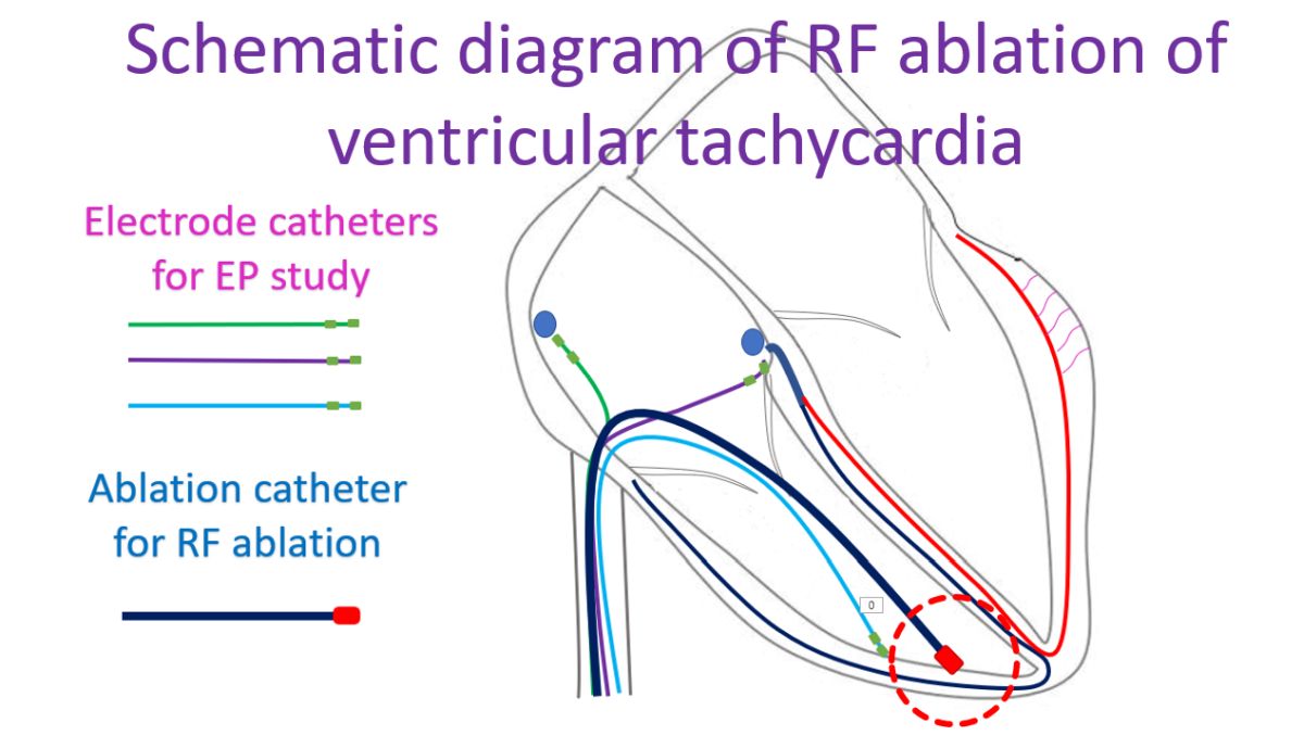
Newer modalities of mapping like 3-D mapping may be more useful in identifying abnormal pathways of electrical activity more accurately prior to RF ablation in difficult cases. Ventricular tachycardia with same morphology of all beats on the ECG, known as monomorphic ventricular tachycardia is the one which is usually targeted for RF ablation. Ventricular tachycardia with multiple shapes known as polymorphic ventricular tachycardia usually needs other forms of treatment like specific medications.
Another important role for RF ablation is in atrial fibrillation, a fast irregular rhythm originating from the upper chambers of the heart. Atrial fibrillation is the commonest sustained heart rhythm abnormality in the population. Success rates for RF ablation are higher in those with episodic atrial fibrillation (paroxysmal atrial fibrillation) than in those with persistent atrial fibrillation. The longer the duration of persistent atrial fibrillation, the lower the likelihood of success. If there is a lot of scarring in the upper chambers due to other diseases, the success rates of RF ablation for atrial fibrillation can be lower.
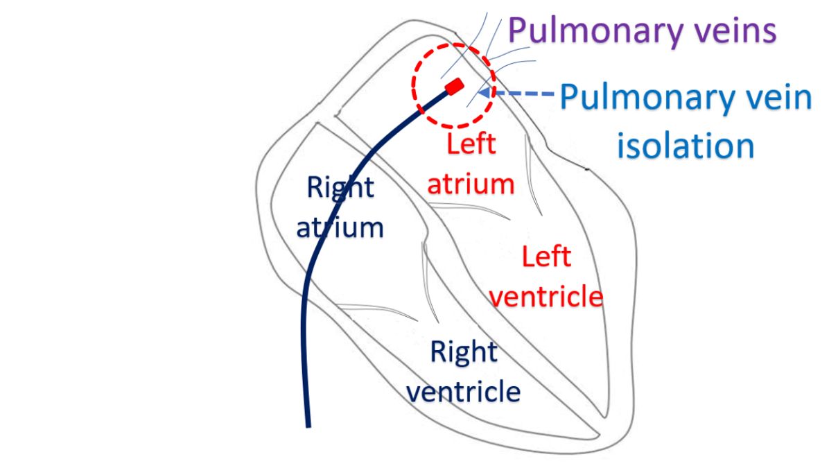
In general, RF ablation for atrial fibrillation is more difficult than that for the usual regular supraventricular tachycardia. This is because more extensive RF ablations may be needed for elimination of atrial fibrillation. An important site for ablation is near the opening of the pulmonary veins into the left atrium. Pulmonary veins drain oxygenated blood from the lungs to the left atrium. Left atrium is the left upper chamber of the heart. The procedure is called pulmonary vein isolation as it isolates the abnormal focus of electrical activity in the pulmonary veins from the rest of the heart.
Related Posts


About The Author
Johnson Francis
Former Professor of Cardiology, Calicut Govt. Medical Kozhikode, Kerala, India. Editor-in-Chief, BMH Medical Journal