What is transposition of great arteries (TGA)?
What is transposition of great arteries (TGA)?
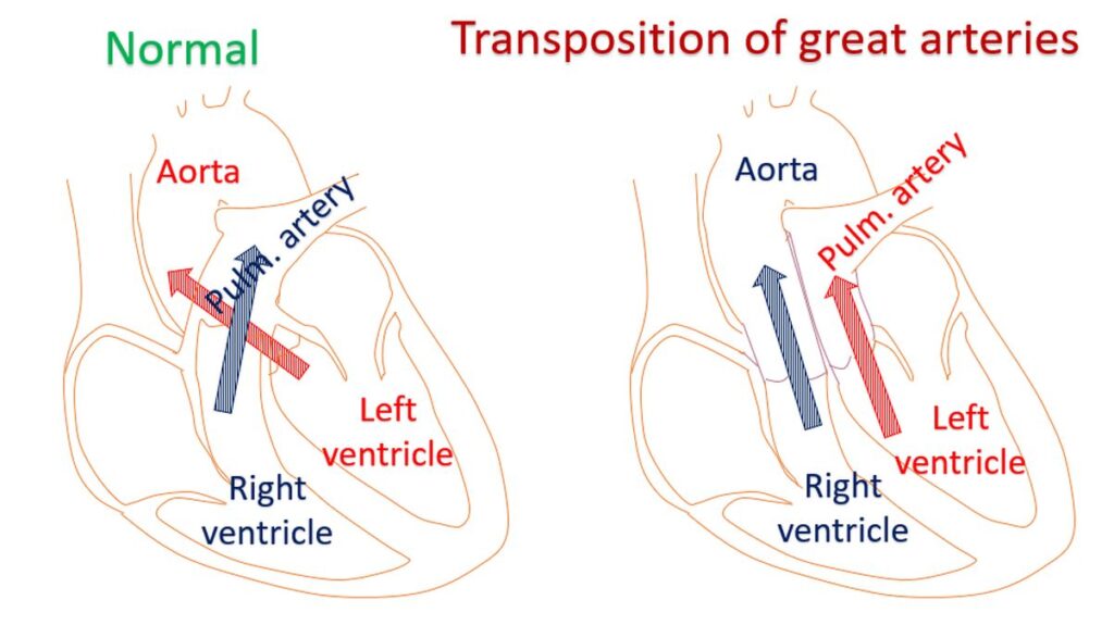
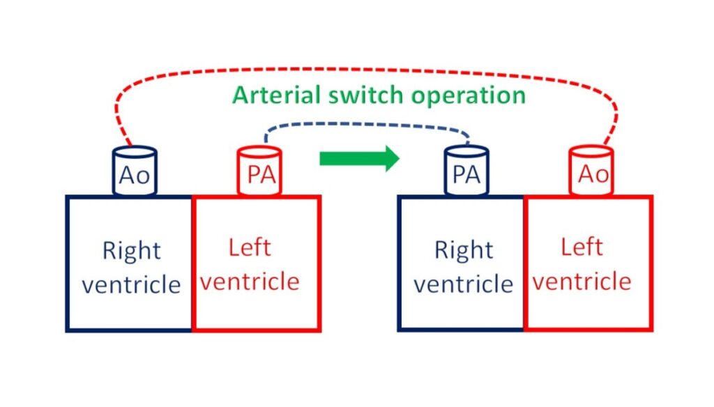
Normally the aorta arises from the left ventricle and pulmonary artery from the right ventricle. In transposition of great arteries, this is reversed so that aorta arises from the right ventricle and pulmonary artery from the left ventricle. Left ventricle is the left lower chamber of the heart and aorta the largest blood vessel carrying oxygenated blood to the whole body. Pulmonary artery carries blood to the lungs from right ventricle for oxygenation.
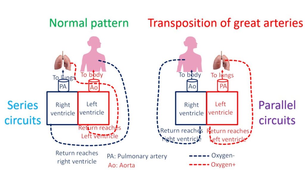
When the aorta is connected to the right ventricle, the blood returning from the body after oxygen extraction is pumped back to the body. There is no chance for blood to get oxygenated from the lungs. Normally the blood returns from the body to the right ventricle and gets pumped to the lungs. It reaches the left ventricle only after oxygenation from the lungs. In transposition this does not happen. So life is impossible in transposition of great arteries unless there is good mixing of the blood within the heart by a defect in the wall between the upper or lower chambers.
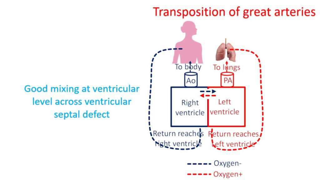
These mandatory defects could be atrial septal defect (ASD) between the upper chambers or a ventricular septal defect (VSD) between the lower chambers. Alternatively a connection between the pulmonary artery and the aorta which is normally there in the baby in the womb (fetus), can persist after birth causing a mixing. This is known as patent ductus arteriosus (PDA). When there is a good mixing, part of the mixed blood goes to lungs for oxygenation and life is possible.
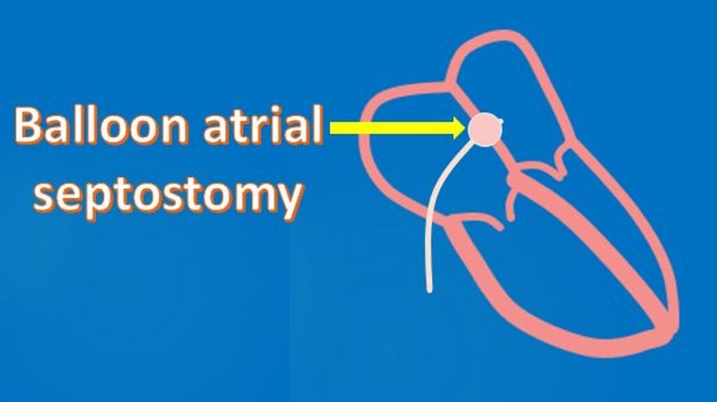
Sometimes a baby may be born with a very small communication between the upper chambers. This defect will not provide sufficient mixing and the baby will be blue due to poor oxygen content of the blood. Emergency treatment given then is to prevent the normal closure of the ductus arteriosus after birth by giving medication. This medication has to be given as a continuous drip. After this emergency treatment, the baby undergoes an emergency procedure known as balloon atrial septostomy. In balloon atrial septostomy, the small opening between the upper chambers is enlarged using a balloon attached to the tip of a thin tube. The tube is usually introduced through the blood vessel in the navel in the if it is done soon after birth. If it gets delayed, blood vessel in the groin may be used as the blood vessel in the navel closes off.
After tiding over the crisis, the baby is planned for an arterial switch operation in which the blood vessels are switched to their normally expected position. That is aorta is disconnected from the right ventricle and connected to the left ventricle. The pulmonary artery is disconnected from the left ventricle and connected to the right ventricle.
This procedure has to be done within about two weeks of birth. For some reason if this is not possible, then several other more complex surgical procedures are planned and executed. The aim of these procedures is to re-route the blood with low oxygen returning from the body to the lungs and the oxygenated blood returning from the lungs to the aorta.
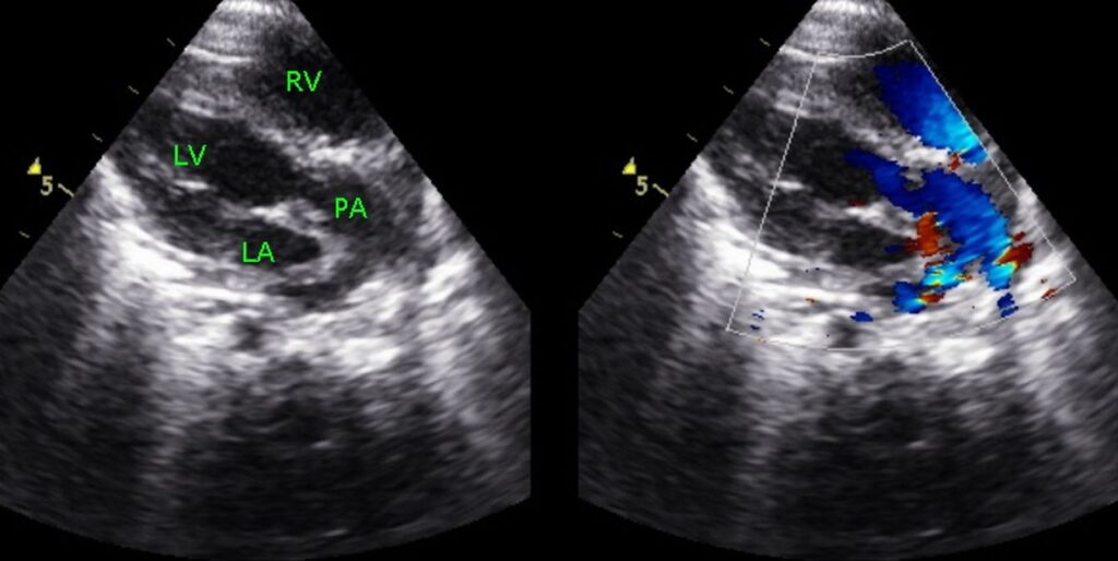
A baby with transposition of great vessels is usually born as a blue baby and develops feature of heart failure soon. Hence it is very important to detect this condition soon after birth. Screening of all newborn babies with pulse oximetry to check oxygen saturation in the blood is useful in detecting such major birth defects of the heart. Confirmation is by an echocardiogram, ultrasound study of the heart. Echocardiogram will also tell about associated defects and help in planning procedures.
Occasionally a baby with a good mixing of blood in the upper or lower chambers of the heart due to an associated ASD or VSD, may present later in life. This is more likely if there is an additional partial obstruction to the blood flow into the lungs, so that lungs are not flooded with blood causing heart failure.
This description is about the more common type of transposition of great arteries. There are other varieties like congenitally corrected transposition of great arteries, which is functionally normal and often missed in younger age and detected much later in life. Multiple birth defects of the heart can also complicate the picture in transposition of great arteries, which can be identified by a meticulously done echocardiogram.
Related Posts


About The Author
Johnson Francis
Former Professor of Cardiology, Calicut Govt. Medical Kozhikode, Kerala, India. Editor-in-Chief, BMH Medical Journal