What is an echocardiogram?
What is an echocardiogram?
Echocardiogram is an ultrasound imaging of the heart. Ultrasound is high frequency sound wave which is not audible to the human ear. The machine sends out ultrasound beams intermittently to the heart and listens for the echo.
Computer synthesised images are then produced from the echoes received from various parts of the heart.
It is similar to the location of aeroplanes in the sky by a radar system. The ultrasound signals are sent and received by a probe placed on the chest in specific locations.
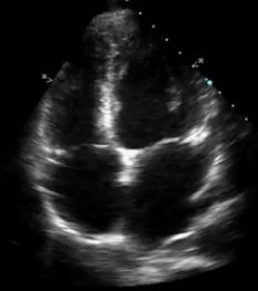
This is an echocardiogram showing all four chambers of the heart and two valves between the upper and lower chambers.
The view is from the lower part of the heart and appears upside down. Here are the two lower muscular chambers – right and left ventricle.
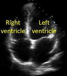
Now the upper chambers – right and left atrium have been marked.
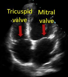
Tricuspid valve on right side and mitral valve on left side have been marked here. Tricuspid valve is between the right atrium and right ventricle and the mitral valve between the left atrium and left ventricle.
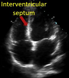
Here the wall between the two lower chambers – interventricular septum has been marked.
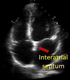
The wall between the two upper chambers is known as the interatrial septum.
Doppler imaging is by analysing the echoes of the ultrasound beam from moving red blood cells. You will be familiar with the difference between the sound of a moving train when it comes towards you and when moves away from you. The same principle is applied in Doppler echocardiography. Signals from blood moving away from the probe is colour coded blue and that moving towards the probe is colour coded red.
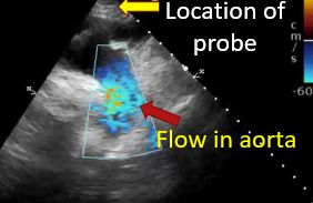
This colour Doppler shows downward movement of blood in the largest blood vessel of the body, known as aorta. As the probe is kept in the upper part here, the downward flow in the aorta is colour coded blue.
Related Posts


About The Author
Johnson Francis
Former Professor of Cardiology, Calicut Govt. Medical Kozhikode, Kerala, India. Editor-in-Chief, BMH Medical Journal