What is aortic stenosis?
What is aortic stenosis?
Aortic stenosis is narrowing of the valve between the left ventricle and aorta. The aortic valve prevents backflow of blood from the aorta into the left ventricle, when it relaxes after a contraction. Left ventricle is the lower muscular chamber of the heart which pumps blood to the whole body. Aorta is the large blood vessel arising from the left ventricle, carrying oxygenated blood.
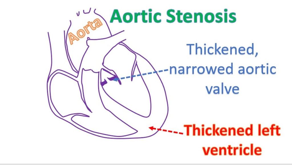
When the aortic valve gets narrowed, the left ventricle finds it difficult to pump blood into the aorta. As the obstruction progresses, the left ventricular muscle becomes thickened, in an effort to compensate for the increased workload. The pressure inside the left ventricle also goes up. A stage is reached when the left ventricle can no longer pump blood well into the aorta when there is severe aortic stenosis. Then the left ventricle starts failing. Initially only the pressure in the left ventricle when it contracts (systolic pressure) increases. When the left ventricle starts failing the pressure inside when it relaxes (diastolic pressure) also rises.
This rise is in turn transmitted to the left atrium the upper chamber from which the left ventricle receives blood in diastole. Rise in left atrial pressure elevates pressure in the pulmonary veins, which carry oxygenated blood to the heart. When the pressure in pulmonary veins (veins of the lungs) go very high, typically above 25 mm Hg, fluid starts collecting within the small air cavities of the lungs (alveoli), where oxygen transfer occurs.
Collection of fluid within the lungs is known as pulmonary edema. The lungs become heavier and breathing becomes difficult. This situation is known as heart failure and is a late symptom of aortic stenosis. When pulmonary edema occurs, lungs are unable to oxygenate the blood properly and oxygen saturation of blood falls.
Another important symptom which can occur in aortic stenosis is chest pain on exertion, known as effort angina. As aortic stenosis progresses, the left ventricular muscle become thickened as a compensatory mechanism in an attempt to overcome the resistance to blood flow. The coronary arteries which supply oxygenated blood to the heart are unable to meet the increased demand of the thickened muscle. This produces chest pain when the person walks or exerts in other ways.
A much more sinister symptom of aortic stenosis is syncope or fainting. In earlier stages it may manifest as dizziness or near syncope on exertion. In severe aortic stenosis, the left ventricular output becomes rather fixed, meaning that it cannot increase much as occurs in a normal heart with exercise. The normal heart can increase its output as much as 6 times at peak exercise. But this cannot occur in severe aortic stenosis. At the same time, peripheral blood vessels dilate during exercise, leading to a fall in blood pressure. This leads to lowering of blood supply to the brain and the person may become transiently unconscious and fall with undue exertion. Very occasionally this can also lead to sudden death.
Aortic stenosis can occur as a long term sequelae of a disease known as rheumatic fever which affects joints and heart valves, more common in developing countries. In developed countries, more common cause is age related degeneration of aortic valve seen in the elderly. In a birth defect known as bicuspid aortic valve, aortic valve has only two leaflets instead of the usual three leaflets. Bicuspid aortic valve can get damaged in the long run and become narrowed, producing aortic stenosis. Very rarely, an infant may be born with only with a single leaflet aortic valve and severe aortic stenosis.
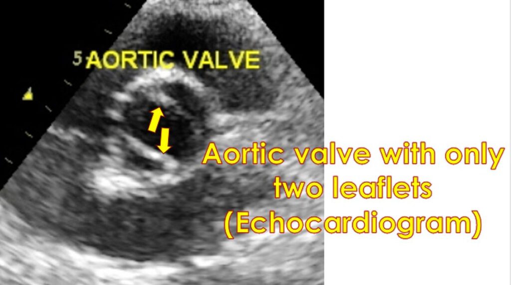
Usual tests done in case of suspected aortic stenosis are ECG, X-ray of the chest and an echocardiogram (ultrasound study of the heart). Those older patients planned for surgery may undergo coronary angiography, an X-ray imaging of the heart after injecting medications into the coronary arteries. Echocardiogram gives an assessment of severity of aortic stenosis and associated thickening of the left ventricle. Measurement of the pumping function of the left ventricle can be done by echocardiography. It will also check whether the valve has a leak in addition. State of the other heart valves are also documented.
Persons with severe aortic stenosis should be careful about exertion due to the risks mentioned earlier. Those in heart failure can be given medications, with limited benefit. Severe aortic stenosis with symptoms needs aortic valve replacement. Usual treatment is replacement with a mechanical or bioprosthetic valve. Those given a mechanical valve needs lifelong medications to prevent clot formation inside the valves. Bioprosthetic valves need such medication for initial few months after the operation.
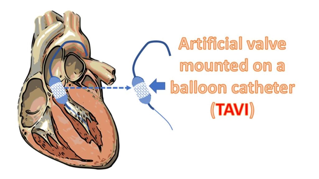
A new, though currently more expensive option is the implantation of an artificial valve through tubes introduced through the groin known as transcatheter aortic valve implantation (TAVI, also known as TAVR – transcatheter aortic valve replacement). It is becoming more popular as it avoids an open heart surgery. Initially it was used in those with high risk for open surgery. But as the techniques improved, TAVI is being used for those at lower risk for surgery also.
Related Posts


About The Author
Johnson Francis
Former Professor of Cardiology, Calicut Govt. Medical Kozhikode, Kerala, India. Editor-in-Chief, BMH Medical Journal