What is WPW syndrome?
What is WPW syndrome?
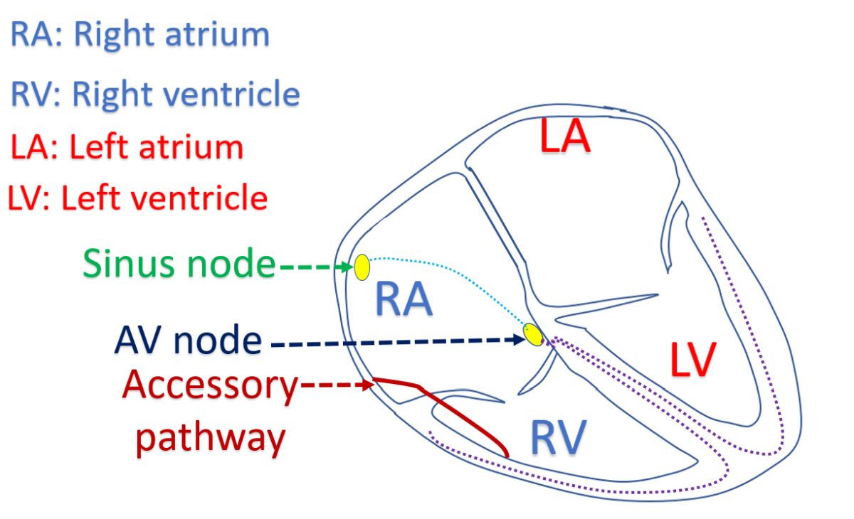
WPW syndrome is short form for Wolff-Parkinson-White syndrome. WPW syndrome is characterized by an accessory electrical conduction pathway from the upper chambers of the heart to the lower chambers causing heart rhythm abnormalities. Heart has a natural pacemaker known as the sinus node situated in the right upper chamber. The electrical signals are conducted to the next relay station at the lower part of that chamber known as the atrioventricular node (AV node). From the AV node, the signals are conducted to the lower chambers after a short delay. This delay is bypassed by the accessory pathway.
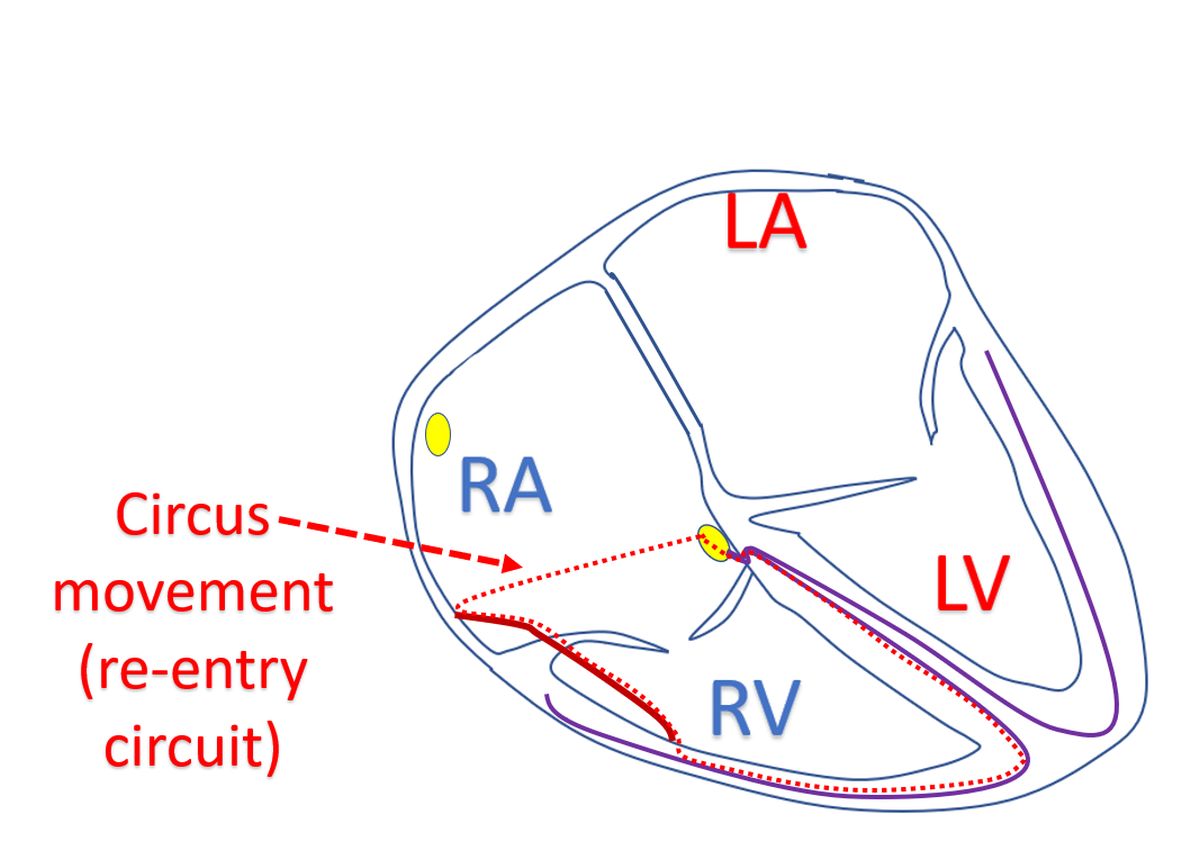
When there are two pathways to the lower chambers – one through the AV node, and another through the accessory pathway, signals can sometimes travel through the pathways in a sequence forming re-entrant circuit. Signals pass down one pathway and return to the upper chambers through the other pathway, forming a circus movement. When the signals pass back and forth through these circuits in a rapid fashion, a very fast heart rate can occur, which is felt by the person as palpitation. If the rate is very fast, heart cannot fill properly between contractions and the amount of blood pumped out by the heart in each contraction comes down.
When the amount of blood pumped out by the heart is low, blood flow to the brain may be affected. Low blood supply to the brain may cause the person to faint when the heart rate is very fast. Sometimes if the fast rate persists for a long period without treatment, it may weaken the heart muscle. This weakening reduces the pumping function of the heart muscle leading to heart failure. In heart failure, there is damming up of blood in the lungs, making it heavier and causing breathlessness.
When a fast rhythm is noted, it can be immediately treated with medications given through the blood vessels of the arms, with monitoring of the heart rhythm, ideally in an intensive care unit. Recurrence can be prevented by giving appropriate medications on a long term basis. Sometimes when a life threatening heart rhythm abnormality is noted, a controlled electrical shock using a device known as defibrillator may have to be given for correcting the heart rhythm.
WPW syndrome can be diagnosed by an ECG, the recording of the electrical activity of the heart. An ultrasound study of the heart known as echocardiogram will check whether there is any associated structural heart disease. An abnormality of the valve between the right upper and lower chambers of the heart known as Ebstein’s anomaly may be associated with WPW syndrome. Location of the accessory pathway can be localized to some extent by noting the pattern in the ECG.
Further localization of the accessory pathway can be made by an electrophysiology study. In electrophysiology study, multiple electrode wires are introduced into the heart through the blood vessels in the groin and neck. Recording of the electrical activity at multiple sites within the heart will localize the abnormal pathway. Once it is localized, it can be removed by a procedure known as radiofrequency catheter ablation. In this procedure, tiny, localized burns are produced in the location of the accessory pathway using very high frequency currents. This removes the electrical conduction in the abnormal pathway and resolves the heart rhythm abnormality.
Related Posts


About The Author
Johnson Francis
Former Professor of Cardiology, Calicut Govt. Medical Kozhikode, Kerala, India. Editor-in-Chief, BMH Medical Journal