What is a PFO?
What is a PFO?
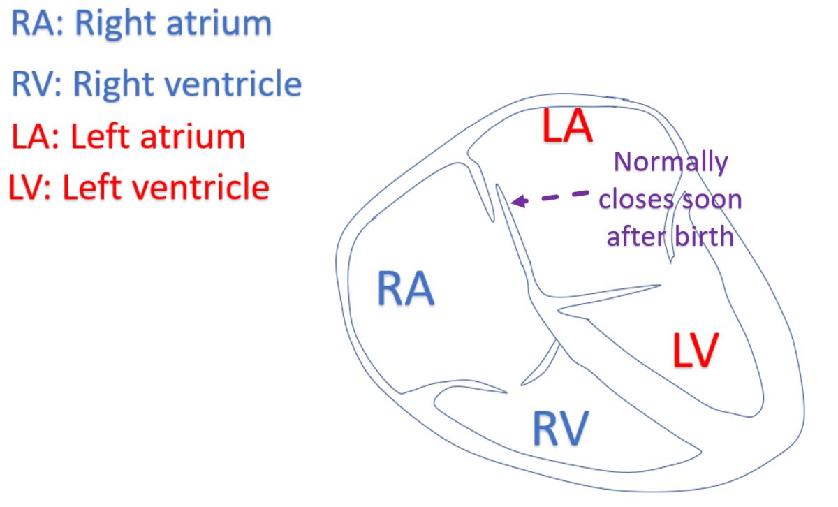
PFO is short form for patent foramen ovale. It is a small communication from right atrium to left atrium. Right atrium is the right upper chamber and left atrium the left upper chamber. Foramen ovale is a communication present in the baby while in the mother’s womb. It closes off soon after birth so that there is no communication between the two upper chambers in most persons. But occasionally, a small opening may persist and then it is called PFO.
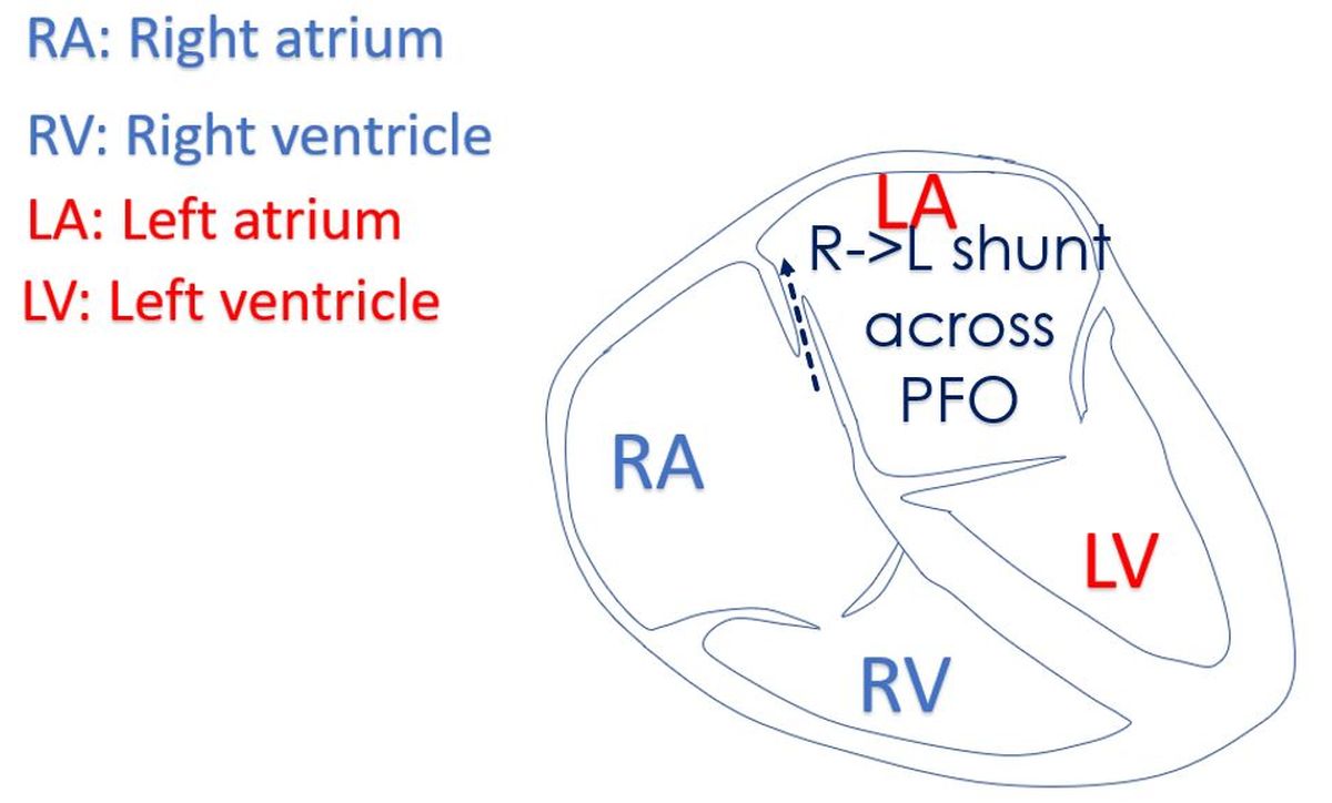
Foramen ovale is an oblique opening through the wall between the two upper chambers of the heart. When the pressure in the left atrium rises after birth as the lungs become functional, it presses on the left side of the opening and closes it. If there is a residual opening, most of the time there is no leakage of blood across the PFO. But occasionally as in straining, the pressure in the right atrium transiently rises so that blood passes from the right atrium to the left atrium across the PFO.
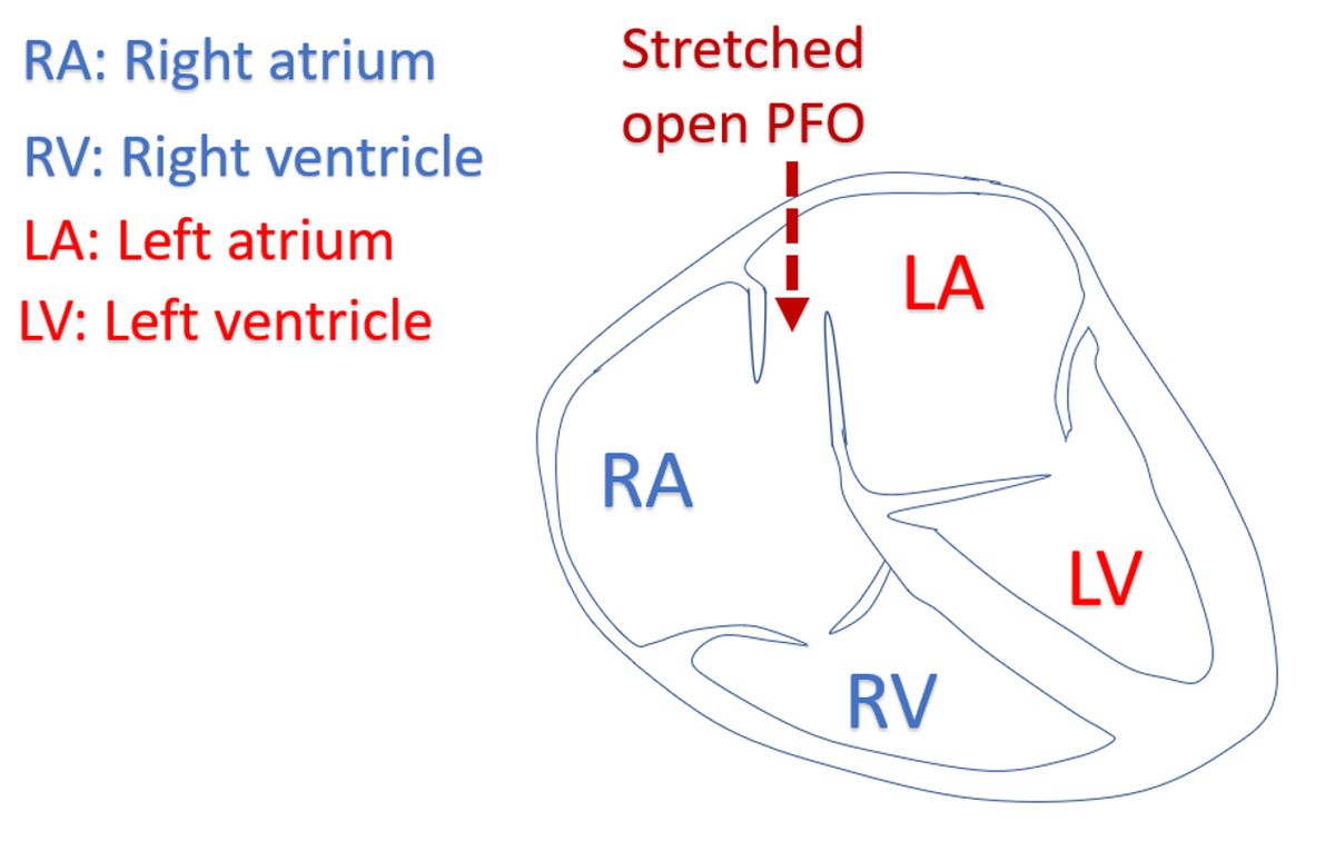
Another situation is a stretched open PFO. When the pressure in the right or left atrium rises significantly due to another disease, the atrium enlarges and stretches the wall between the two atria along with it. When the PFO is stretched, blood can flow either way, depending on which side has the higher pressure. If it is the left atrium which is enlarged, the stretched open PFO will shunt blood in a left to right direction and vice versa. If too much of right atrial blood with lower oxygen content reaches the left atrium, the oxygen saturation in the blood vessels of the body can fall, leading to bluish discoloration of skin.
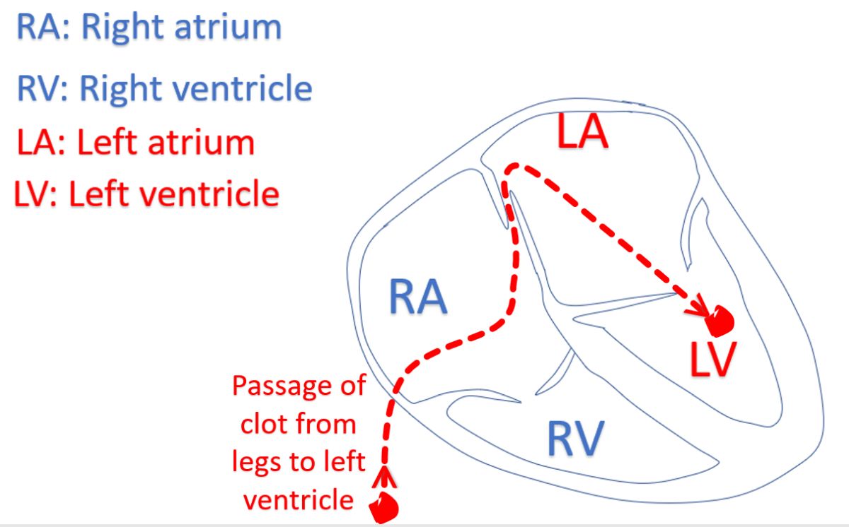
Small PFO cannot produce much problem due to the shunting of blood across it unless it is stretched open by other diseases. But small clots from the leg veins or tummy can occasionally pass across it to the left atrium. This is a risky situation. The clot can move from the left atrium to the left ventricle and then to the aorta. Left ventricle is the lower muscular chamber which pumps blood to the whole body. Aorta is the large blood vessel arising from the left ventricle. From the aorta the clot can move to any part of the blood circulation. If it gets lodged in a blood vessel of the brain and blocks it, a stroke with paralysis may occur.
Passage of blood clots from the right side of the heart to the left side across a PFO is known as paradoxical embolism. Most important problem with paradoxical embolism is stroke, though it can also get lodged in any other blood vessel of the body and cause damage to that region. Some advocate closure of the PFO with a device, especially after a stroke, to prevent recurrence. This is considered only if the right to left shunting across the PFO is demonstrated.
PFO can be documented by ultrasound study of the heart known as echocardiogram. It will also show right to left shunting during certain types of strain, suggesting the risk of paradoxical embolism. Another test is transcranial Doppler, an ultrasound study of the head which looks for tiny air bubbles in the blood vessels of the brain. For detecting a right to left shunt across the PFO, agitated saline containing tiny air bubbles is injected into the forearm vein. If the air bubbles are detected by the transcranial Doppler machine, it is presumed that tiny blood clots can also pass across the PFO to produce a stroke. Right to left flow of tiny air bubbles in the agitated saline will be seen in the left atrium on echocardiogram.

Related Posts


About The Author
Johnson Francis
Former Professor of Cardiology, Calicut Govt. Medical Kozhikode, Kerala, India. Editor-in-Chief, BMH Medical Journal