What is an EP study?
What is an EP study?
EP study is short form for electrophysiology study. It is a test done to assess the electrical system of the heart. Heart has an electrical system in which regular pulses are produced from the right upper chamber, by a small structure known as the sinus node. Sinus node is the natural pacemaker of the heart. The signals are conducted down the upper chambers to the atrioventricular (AV node) situated at the junction between the upper and lower chambers.
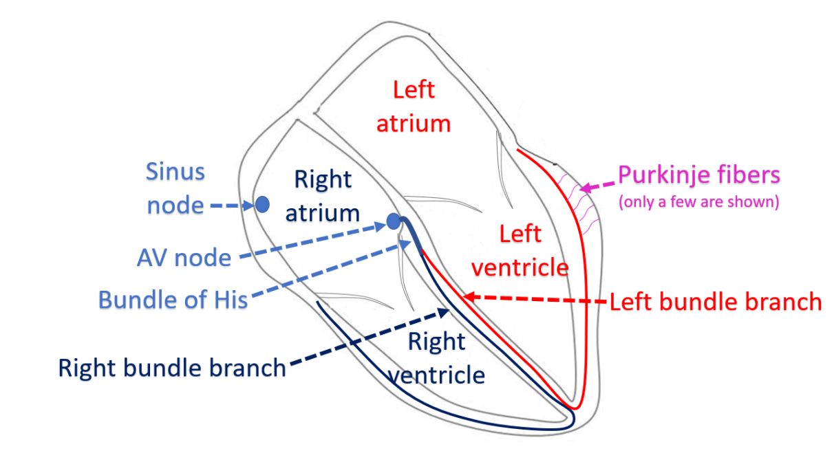
From the AV node, the signals are conducted to the lower chambers through the bundle of His and its branches. The right and left lower chambers receive one branch each, right and left bundle branches. These further divide into minute conduction fibers known as Purkinje fibers, which reach each and every heart muscle cell. Thus the heart is able to work in a regular sequence by the control of the electrical system of the heart.
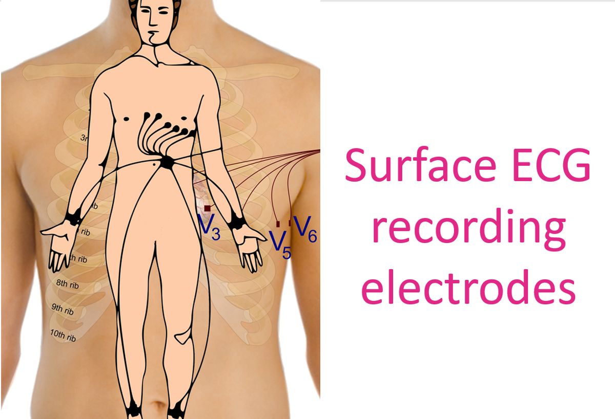
When the electrical system of the heart is diseased, several heart rhythm disorders can occur, which could be fast rhythms, slow rhythms and irregular rhythms. These rhythms are usually picked up by the electrical recording of the heart known as ECG. ECG uses several electrodes placed on the body surface in specified locations. Hence the usual ECG is also called surface ECG.
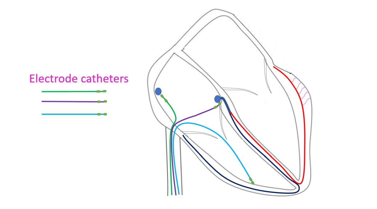
When the signals travel from the heart to body surface, the signal strengths come down and there is mixing of signals from different parts of the heart. So finer details may not be available from a surface ECG. If signals are recorded directly from the site of origin within the heart, the signals are stronger and more accurate with less mixing by signals from other parts. This is done during electrophysiology study (EP study).
In EP study, small wires with multiple electrodes (electrical contacts) on them are introduced into the heart through the blood vessels of the neck and groin. This is done under local anaesthesia through small holes made in the skin. The wires are guided into the heart using X-ray imaging equipment in an electrophysiology laboratory (EP lab). Electrical signals from specified regions of the heart are recorded using a high fidelity recorder (EP recorder).
While the surface ECG has only a limited number of channels, typical 12 leads for usual recordings, EP study uses a large number of electrodes and channels. Number of channels increases when highly specialized studies are needed. The recording speed is also much higher than in surface ECG. While surface ECG recordings are at 25 mm/s, EP tracings are taken at 100 or 200 mm/s. They are also amplified as needed to get finer details, by the EP recorder.
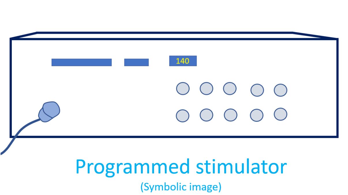
Another device used in the EP lab is a programmed stimulator. The stimulator gives out signals to the heart at specified intervals and checks the response of the heart and how the signals are conducted to various parts of the heart. Stimulator can give signals at much higher rate than the usual heart beats. In this way it is possible to find out how the heart responds to fast rates, even though the person is at rest in the EP lab.
Mapping out the response to the signals given by the programmed stimulator as well the natural signals of the heart are an important part of the EP study. From the pattern of spread of the signals within the heart, it is possible to find out the mechanism of the heart rhythm abnormality noted in the surface ECG. This is very useful in those with fast heart rhythms, slow rhythms and irregular rhythms. Once the exact mechanism of the heart rhythm abnormality is found, more accurate treatment decisions can be made.
Related Posts


About The Author
Johnson Francis
Former Professor of Cardiology, Calicut Govt. Medical Kozhikode, Kerala, India. Editor-in-Chief, BMH Medical Journal