ECG Basics – A Brief Review >>Twelve Lead ECG
Most commonly recorded ECG is a 12 lead ECG. Twelve leads in a standard ECG are as follows:
- Standard Limb Leads: I, II, III
- Augmented Limb Leads: aVR, aVL, aVF
- Chest Leads: V1, V2, V3, V4, V5, V6
Thus a standard 12 lead ECG does not include right chest leads known as V3R, V4R, V5R etc. But in most cases 12 lead ECG includes a long rhythm strip – either lead II or V1 or both, for facilitating rhythm analysis.
If the ECG machine has only a single recording channel, 12 leads are recorded sequentially. More sophisticated recorders have facility to record 3 or even 12 channels simultaneously. Simultaneous multichannel recording can record all leads of a single beat and is better for analysis of complex cardiac rhythm abnormalities.
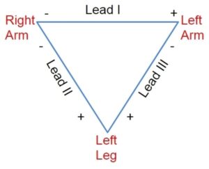
Electrode combination for limb leads:
Lead I: Left arm positive, Right arm negative
Lead II: Right arm negative, Left foot positive
Lead III: Left arm negative, Left foot positive
Derivation of augmented limb leads:
Augmented limb leads are derived using the limb lead electrode potentials. All the three limb leads are internally connected to a central terminal (Wilson’s central terminal) using fixed value resistors. Usual limb lead recordings are called bipolar recordings with one limb serving as positive electrode and another as negative electrode as illustrated above. Unipolar recordings are obtained by taking the limb electrode as positive and central terminal as negative. Unipolar limb leads are called VR, VL and VF depending on whether the positive electrode is at right arm, left arm or left leg. Voltages of these recordings can be augmented by disconnecting the connection to the corresponding limb electrode from the central terminal, causing almost 50% increase in the amplitude of the recordings. The augmented leads thus generated are called aVR, aVL and aVF.
Electrode positions for chest leads:
Chest electrodes are placed on the anterior chest wall as follows:
V1: 4th right intercostal space, right sternal edge
V2: 4th left intercostal space, left sternal edge
V3: Midway between V2 and V4
V4: 5th left intercostal space, midclavicular line
V5: Anterior axillary line, same horizontal line as V4
V6: Mid axillary line, same horizontal level as V4
Chest leads are often positioned at varying positions by different operators due to difficulty in ascertaining intercostal spaces accurately in obese patients and females. This may cause difficulty in comparing serial ECGs. Hence every effort should be made to place the electrodes accurately. In some situations when accurate comparison of serial ECGs are needed, marking electrode position with a skin marker pen may be needed.
Chest leads are named C1, C2, C3, C4 etc. by some manufacturers. Here ‘C’ would stand for ‘chest’. In the conventional naming system ‘V’ stands for ‘voltage’.
Colour coding for electrodes
Conventionally, right arm electrode was given a colour coding of red, left arm electrode yellow, left leg electrode green and the neutral electrode in right leg was given a black colour. But current manufacturers sometimes use different colour coding systems so that one has to verify the letters written on the electrodes before using them – RA for right arm, LA for left arm, LL for left leg and RL for right leg. Moreover, a previous user might have changed the position of the clips attached to electrodes so that the colour coding can be misleading. Hence it is essential to make sure of the lead connections to the ECG machine before recording the ECG to avoid wrong lead placements, which is the commonest technical error in ECG recording.
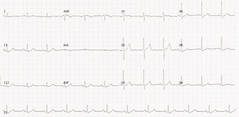
12 lead ECG showing normal pattern of P, QRS and T waves