Choosing an antihypertensive medication in the outpatient department (OPD) relies on a stepwise approach driven by the patient’s baseline blood pressure, compelling comorbidities, and demographic factors, aligning with
In a busy cardiac catheterization lab, operators and support staff are exposed to scatter X-ray radiation for extended periods. While traditional protective aprons use pure lead, modern lead-free
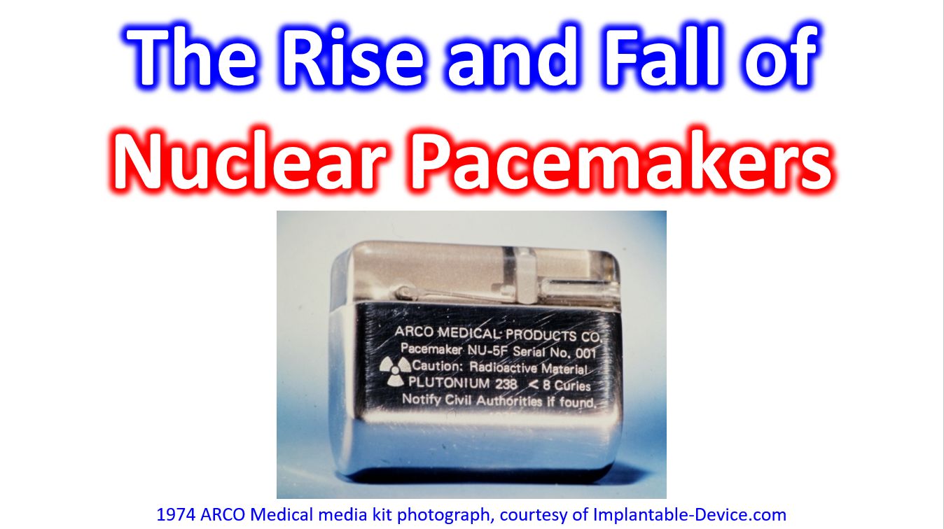
In the late 1960s and 1970s, nuclear-powered pacemakers fueled by Plutonium-238 were hailed as a medical marvel. They offered incredible longevity, with power supplies capable of lasting 20
The Syncope-Stopper Study (ANZCTR Registration: ACTRN12622001553763) is an Australian randomized controlled trial evaluating the safety, efficacy, and cost-effectiveness of early pacemaker implantation compared to standard care for high-risk
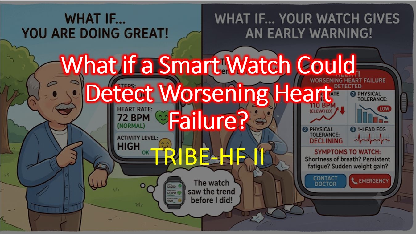
Here is a comprehensive review of the TRIBE-HF II study, which explores the intersection of wearable technology and heart failure management. Overview of TRIBE-HF II The TRIBE-HF II
The Fifth Universal Definition of Myocardial Infarction (UDMI) was officially published on August 28, 2026, as a joint consensus document by the European Society of Cardiology (ESC), the
The “Pickelhaube sign” (also referred to as the Spiked Helmet Sign) refers to two entirely distinct clinical entities depending on whether you are looking at an echocardiogram or
The Dunning classification is specifically used to grade iatrogenic aortocoronary dissection (IACD), a rare but severe complication that occurs when an intimal tear during percutaneous coronary intervention (PCI)
While both concepts are named after the renowned French electrophysiologist Philippe Coumel, they refer to fundamentally different aspects of cardiology. Coumel’s sign is a specific diagnostic electrocardiographic finding
While both are named after the same person, the Aslanger Pattern and Aslanger Sign describe entirely different phenomena on a 12-lead ECG. One is a critical, life-threatening ischemic