Echo Quiz – Discussion
Echo Quiz – Discussion
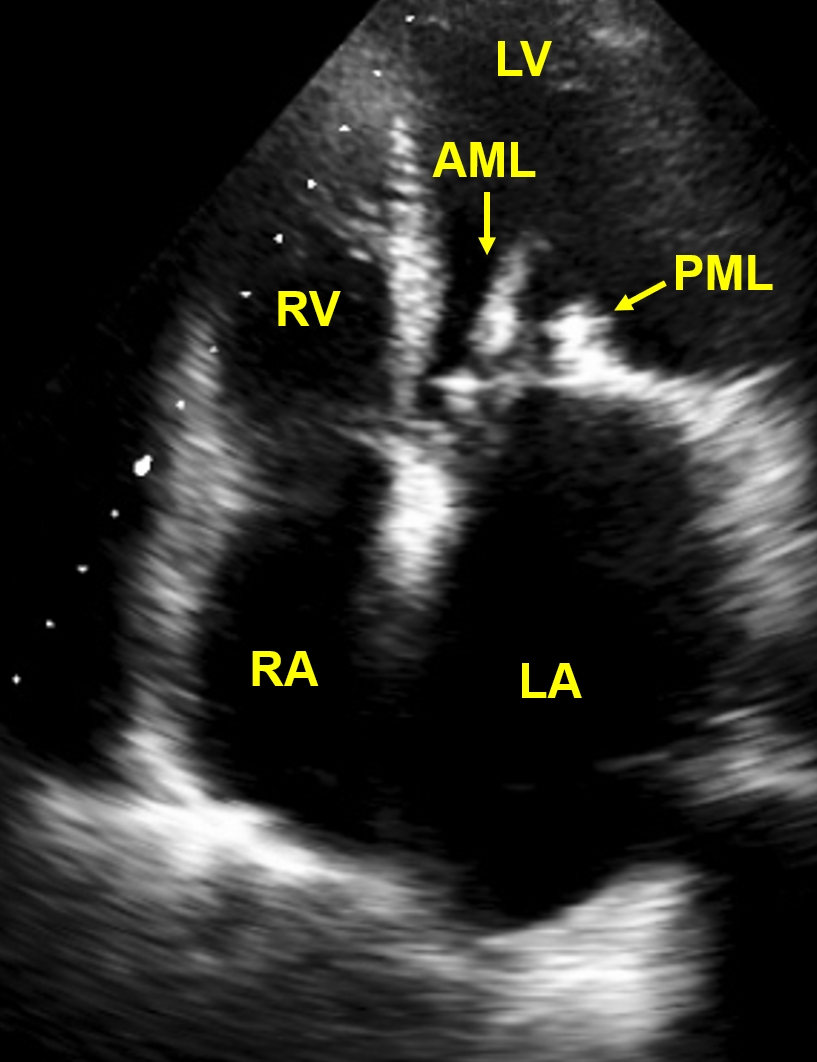
RA: Right atrium; LA: Left atrium; RV: Right ventricle; LV: Left ventricle; AML: Anterior mitral leaflet; PML: Posterior mitral leaflet
This echocardiogram in apical four chamber view shows dilated left atrium and thickened mitral leaflets. Diastolic separation of mitral leaflets is reduced, indicating mitral stenosis. Regions of calcification are visible in both mitral leaflets. Such densely fibrotic and calcified mitral leaflets are seen rheumatic mitral stenosis. Left atrium is dilated due to the hemodynamic load introduced by poor emptying in diastole. Thrombi can form in the dilated left atrium with stasis in mitral stenosis and lead to systemic embolism.
Related Posts


About The Author
Johnson Francis
Former Professor of Cardiology, Calicut Govt. Medical Kozhikode, Kerala, India. Editor-in-Chief, BMH Medical Journal