Bileaflet prosthetic mitral valve – Fluoroscopy video
Bileaflet prosthetic mitral valve – Fluoroscopy video video with narration
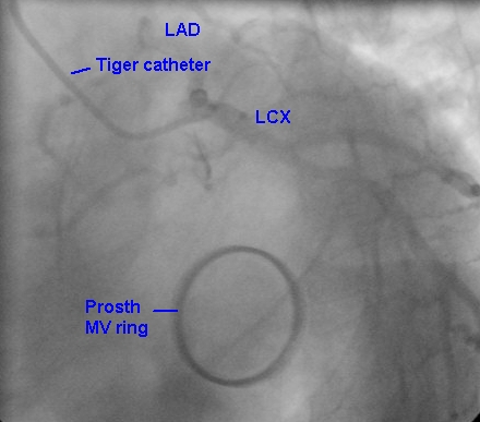
Prosthetic mitral valve ring is seen in the lower part of the image. The leaflets are not visible as they are in the closed position. There is a vague circular outline within the sewing ring, which is much more radio opaque than the leaflets. Upper part of the fluoroscopic image shows the Tiger trans radial catheter in the left coronary artery. The left main coronary artery seems to be short and bifurcates into left anterior descending coronary artery (LAD) and left circumflex coronary artery (LCX). LAD is seen coursing upwards initially in this left anterior oblique (LAO) caudal view. This view is also called the ‘spider view’ as the coronary tree resembles the legs of the spider in this view. LCX gives off a large obtuse marginal (OM) branch. The vessels are not clear in this view as the frame has been frozen in a position to show the valve ring without concentrating on the vascular tree.
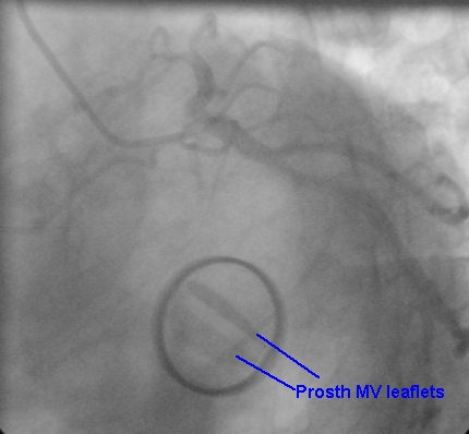
In this view the prosthetic leaflets are partially open so that the leaflets are seen well. The two leaflets have different shape because the view is not exactly en-face to the leaflets, one being seen more tilted than the other. The gap between the leaflets in diastole is seen well. Such good movement excludes the possibility of a stuck valve. Hence fluoroscopy is one of the good ways to establish the movements of prosthetic leaflets. Such a clear view can never be obtained by echocardiography due to acoustic shadowing, which is quite severe with metallic prosthesis.
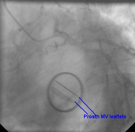
Prosthetic mitral leaflets seen in fully open position, almost perpendicular to the imaging plane. One leaflet is seen just as line as it is exactly perpendicular to the image plane. The other leaflet is seen with larger size because it is partly oblique to the imaging plane. The other leaflet is seen with larger size because it is partly oblique to the imaging plane. Left coronary angiogram in the LAO caudal view is seen in the upper part of the image.
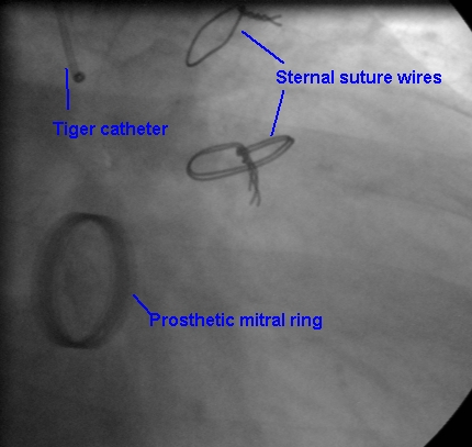
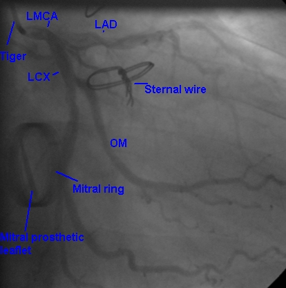
Prosthetic mitral valve ring seen on fluoroscopy in the RAO caudal view. Two sternal suture wires are seen in the upper part of the image. The Tiger catheter is seen engaging the left coronary ostium. Valve leaflet (tilting disc) is not seen clearly in this image.
This frame shows the prosthetic mitral ring along with one leaflet at an angle. The Tiger catheter tip is seen in the left main coronary artery (LMCA). LAD: left anterior descending coronary artery; LCX: left circumflex coronary artery; OM: obtuse marginal branch of LCX. There are two more obtuse marginal branches originating below the marked OM. The terminal LCX is a tiny vessel which is seen faintly.
Related Posts

About The Author
Johnson Francis
Former Professor of Cardiology, Calicut Govt. Medical Kozhikode, Kerala, India. Editor-in-Chief, BMH Medical Journal