Cardiac CT – Pulmonary veins and left atrium
Cardiac CT – Pulmonary veins and left atrium
 Cardiac CT scan showing pulmonary veins and left atrium
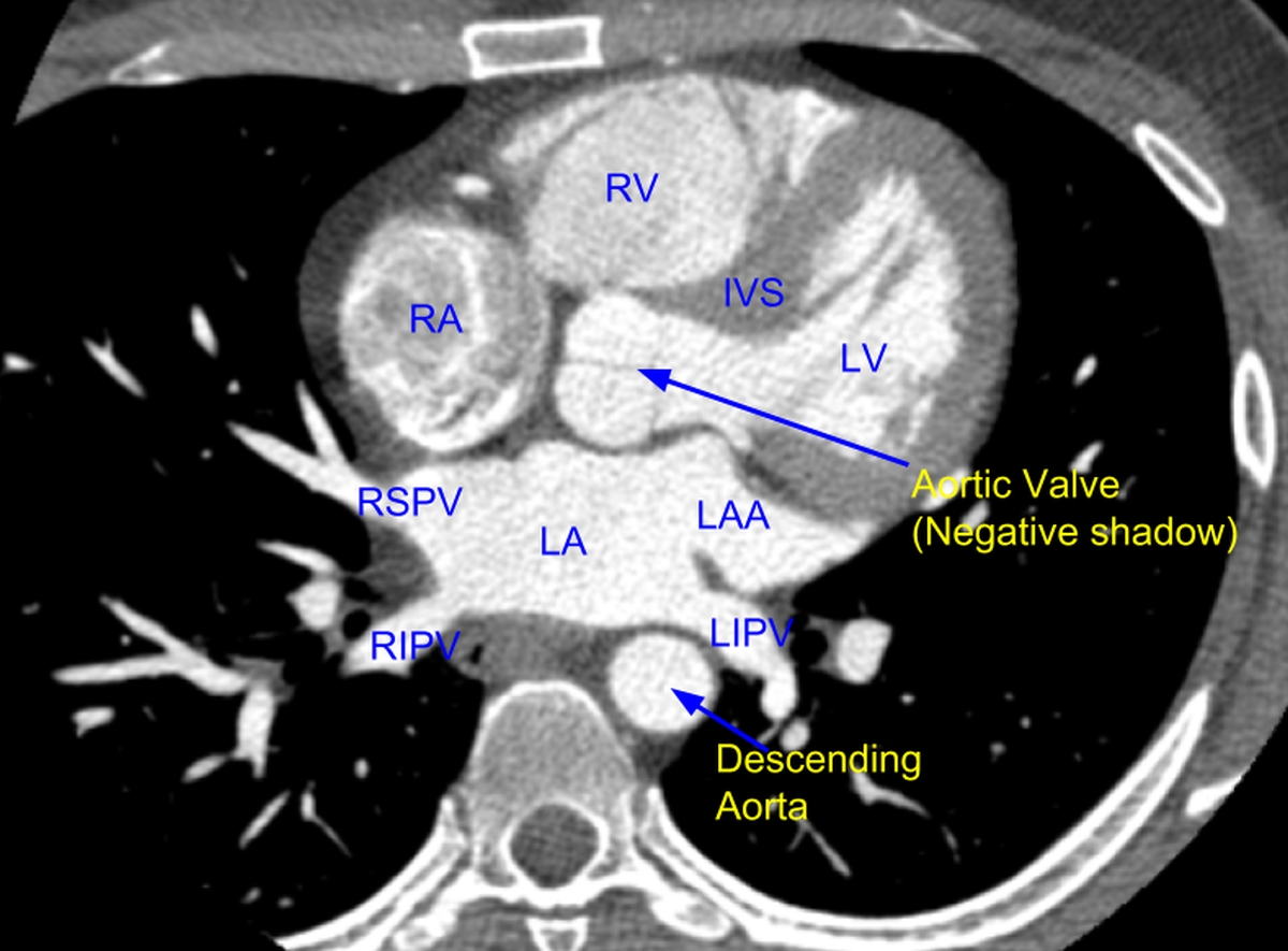
Cardiac CT scan at the level where pulmonary veins join the left atrium:
RSPV: Right superior pulmonary vein; LA: Left atrium; LAA: Left atrial appendage; RIPV: Right inferior pulmonary vein; LIPV: Left inferior pulmonary vein. RA: Right atrium; RV: Right ventricle; LV: Left ventricle. IVS: Interventricular septum. Left ventricle is connecting to the aorta through a narrowed region which is the left ventricular outflow tract (LVOT – not labelled). Part of the blue arrow pointing to the aortic valve is through the LVOT. Aortic leaflets are seen as negative shadows within the contrast filled aorta. Since it is not a true cross sectional image of the aorta, the aortic leaflets take an inverted T pattern. In a true cross section it would be seen as a tri-radiate pattern. Interventricular septum (IVS) is also seen as a negative shadow between contrast filled right and left ventricular cavities. In this image it can be appreciated that the right ventricle (RV) is an anterior chamber placed substernaly while the left ventricle (LV) has a more posterior location. Contrast density is more on the left sided chambers as it is a delayed image after the intravenous contrast injection.
Updated on 1st May 2020
Multidetector computed tomography (MDCT) is an excellent imaging modality to study pulmonary veins prior to pulmonary vein ablation in atrial fibrillation. It is also useful to observe for significant sequelae like pulmonary vein stenosis post procedure [1].
Reference
- Cronin P, Sneider MB, Kazerooni EA, Kelly AM, Scharf C, Oral H, Morady F. MDCT of the left atrium and pulmonary veins in planning radiofrequency ablation for atrial fibrillation: a how-to guide. AJR Am J Roentgenol. 2004 Sep;183(3):767-78.
Related Posts


About The Author
Johnson Francis
Former Professor of Cardiology, Calicut Govt. Medical Kozhikode, Kerala, India. Editor-in-Chief, BMH Medical Journal