Prosthetic valve seen on this X-ray is:
a) Starr-Edwards prosthetic aortic valve
b) Starr-Edwards prosthetic mitral valve
c) Starr-Edwards prosthetic pulmonary valve
d) Starr-Edwards prosthetic tricuspid valve
ECG pattern is diagnostic of: a) Right bundle branch block b) Anterior wall myocardial infarction c) Brugada syndrome d) Early repolarization syndrome Correct answer
Left anterior hemiblock is characterized by all of the following except: a) Left axis deviation b) rS complexes in II, III, aVF c) No significant QRS widening d)
AF (atrial fibrillation) can lead on to VF (ventricular fibrillation) in: a) Catecholaminergic polymorphic ventricular tachycardia b) Andersen syndrome c) WPW syndrome d) None of the above Correct
Gene mutations described in CPVT (Catecholaminergic Polymorphic Ventricular Tachycardia) include all of the following except: a) Sodium channel b) Cardiac ryanodine receptor (RyR2) c) Calsequestrin d) Ankyrin B Correct
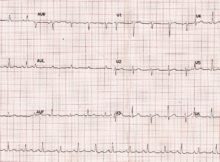
(Click on the image for an enlarged view) ECG finding is suggestive of: a) Myocardial infarction b) Dilated cardiomyopathy c) Pericardial effusion d) None of the above Correct
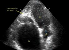
Diagnosis based on the echocardiogram: a) Ebstein’s anomaly of tricuspid valve b) Idiopathic dilatation of right atrium c) Endomyocardial fibrosis d) None of the above Correct Answer