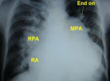
This chest X-ray is suggestive of: a) Primary pulmonary hypertension b) Atrial septal defect with pulmonary hypertension c) Ventricular septal defect with pulmonary hypertension d) Idiopathic dilatation of
Giant wide T inversion may be seen in all except: a) After a cardiac arrest b) Subarachnoid hemorrhage c) Hyperkalemia d) Takotsubo cardiomyopathy Correct answer: c) Hyperkalemia Giant T wave
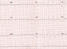
This ECG shows: (Click on the image for an enlarged view) a) Anterior wall infarction b) Anterior wall infarction with right bundle branch block c) Anterior wall infarction
Type II truncus arteriosus is: a) Common pulmonary trunk arises from the truncus arteriosus and divides into left and right pulmonary arteries b) Two pulmonary arteries arise separately
Which of the following is not true of Mahaim fibre tachycardia? a) Left bundle branch block pattern b) Normal baseline ECG c) Mahaim fibres do not conduct retrogradely d)
Wide QRS tachycardia at 300 per minute can occur with: a) Atrial fibrillation with Wolff–Parkinson–White syndrome (WPW) syndrome b) Supraventricular tachycardia with aberrancy in the very young c) Atrial flutter
May-Thurner syndrome is: a) Chromosomal defect b) Iliac vein compression by the crossing iliac artery c) Coarctation of aorta with cerebral aneurysms d) Pulmonary stenosis with webbing of
Inferior vena caval filter is deployed: a) Above the renal veins b) At the renal veins c) Below the renal veins d) Below the hepatic veins Correct answer: c)
Potential complication/s of bilateral internal iliac occlusion due to a stent graft: a) Gluteal claudication b) Erectile dysfunction c) Bowel ischemia d) All of the above Correct answer: d) All
Swiss cheese VSD is: a) Sub-pulmonic VSD b) Peri-membraneous VSD c) Multiple muscular VSD d) None of the above Correct answer: c) Multiple muscular VSD Multiple muscular ventricular septal