Coarctation of aorta – angiogram
Coarctation of aorta – angiogram
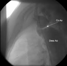
Still picture from an aortogram in coarctation of aorta in lateral view. The catheter is seen along the anterior border of the aorta. Prestenotic (above) and post stenotic dilatation of aorta are visible. Post stenotic dilatation is much more prominent. In this case, though the gradient across the defect was low the post stenotic dilatation was considerable. This illustrates the fact that there is no direct correlation between severity of the obstruction and the post stenotic dilatation. The coarct segment is seen as a shelf like negative shadow extending anteriorly from the posterior border of the aorta (arrow marked Co A). The region marked as Desc Ao is the post stenotic dilatation. Left subclavian artery is seen originating just above the coarctation and coursing upwards. So this is a classical post subclavian juxtaductal coarctation of aorta. LAO view of the same case is given below.
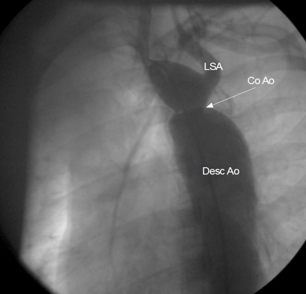
Coarctation of aorta in LAO viewThis is a still picture from aortogram of coarctation of aorta in LAO view. LSA: left subclavian artery. Coarct segment is seen as a constriction just below the origin of the left subclavian artery. The prominent post stenotic dilatation of descending aorta is also visible. Prestenotic dilatation is not very prominent.
Related Posts

About The Author
Johnson Francis
Former Professor of Cardiology, Calicut Govt. Medical Kozhikode, Kerala, India. Editor-in-Chief, BMH Medical Journal