Cor triatriatum sinister and dexter
Cor triatriatum sinister and dexter
Abstract: Cor triatriatum sinister and dexter: Cor triatriatum or heart with three atrial chambers, could be either cor triatriatum sinistrum or cor triatriatum dexter depending on which side the additional chamber is present.
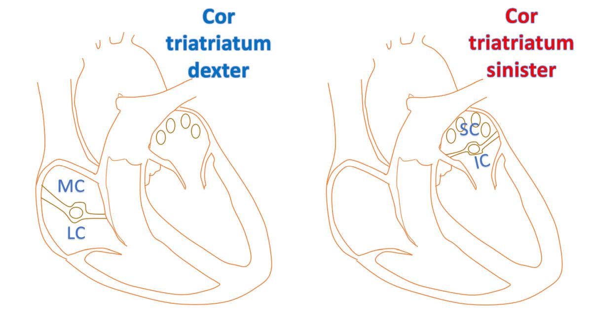
Cor triatriatum sinister and dexter: In cor triatriatum (cor triatriatum sinister [1]) there is a superior and inferior chamber with a membrane dividing the left atrium. In cor triatriatum dexter [2], right atrium is divided into a medial and a lateral chamber. Though both are rare congenital anomalies of the heart, between the two, cor triatriatum dexter is less common.
Cor triatriatum sinister (or sinisterism)
Cor triatriatum sinister is often associated with other cardiac malformations, in up to eighty percent of the cases, with secundum atrial septal defect and anomalous pulmonary venous connection being the most common associated anomalies. The superior chamber receives the pulmonary veins and the inferior chamber is connected to the atrioventricular valve. The inferior chamber also has the atrial appendage and true interatrial septum. Hence it is the true atrium. Size and number of communications between the upper and lower chambers are variable. Sometimes the accessory chamber may connect with the right atrium.
Symptoms depend on whether the connection between the two chambers is obstructive or not. If it is obstructive symptoms are likely due to pulmonary venous hypertension. Symptoms could also be due to other associated cardiac malformations. If there is no significant obstruction, it may be an incidental finding at echocardiography done for other reasons. In that case no treatment is required. Surgical correction can be done if the membrane between the two chambers is obstructive. Three dimensional echocardiography can be useful in planning surgical correction as it gives a good spatial orientation of the chambers and the dividing structure [3].
Cor triatriatum dexter
In cor triatriatum dexter, the right atrium is divided into two chambers possibly by the persistent right sinus venosus valve.4 It is a combination of the fetal Eustachian valve which guards the inferior vena caval orifice and the Thebesian valve which guards the coronary sinus ostium. In the fetal life it is meant for directing the oxygenated blood coming from the umbilical vein through the inferior vena cava to the left atrium across the foramen ovale.
A non obstructive septum is often an incidental finding while an obstructive partition can cause features of right heart failure with systemic venous congestion.
References
- Nassar PN, Hamdan RH. Cor Triatriatum Sinistrum: Classification and Imaging Modalities. Eur J Cardiovasc Med. 2011 Jan;1(3):84-87.
- Yarrabolu TR, Simpson L, Virani SS, Arora H, Navarijo J, Stainback RF. Cor triatriatum dexter. Tex Heart Inst J. 2007;34(3):383-5.
- Jacobs A, Weinert LC, Goonewardena S, Gomberg-Maitland M, Lang RM. Three-dimensional transthoracic echocardiography to evaluate cor triatriatum in the adult. J Am Soc Echocardiogr. 2006 Apr;19(4):468.e1-4.
- Hansing CE, Young WP, Rowe GG. Cor triatriatum dexter. Persistent right sinus venosus valve. Am J Cardiol. 1972 Oct;30(5):559-64.
Related Posts


About The Author
Johnson Francis
Former Professor of Cardiology, Calicut Govt. Medical Kozhikode, Kerala, India. Editor-in-Chief, BMH Medical Journal