D shaped LV cavity in severe RVH
D shaped LV cavity in severe RVH
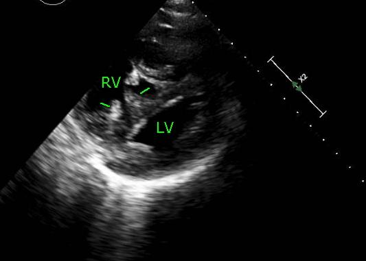
Echo from parasternal short axis view shows a D shaped LV cavity (left ventricular cavity) alongside a grossly hypertrophied right ventricle (RV).
Normally the left ventricular cavity has a circular shape in parasternal short axis view. When there is right ventricular volume overload, the interventricular septum bulges towards the left ventricle and the left ventricular cavity assumes a D shape in diastole. But as the left ventricle (LV) contracts in systole, the pressure rises above that of right ventricle and the LV assumes a circular shape.
When there is severe pulmonary hypertension associated with elevated right ventricular systolic pressures, the interventricular septum bulges into the LV in both systole and diastole so that LV cavity has a D shape through out. In the echo image shown, right ventricular trabeculae can be seen as grossly hypertrophied (green markers). See also: Eccentricity index for right ventricular overload, an echocardiographic index for separation of right ventricular volume and pressure overload [1].
Reference
- Ryan T, Petrovic O, Dillon JC, Feigenbaum H, Conley MJ, Armstrong WF. An echocardiographic index for separation of right ventricular volume and pressure overload. J Am Coll Cardiol, 1985; 5:918-927.
Related Posts

About The Author
Johnson Francis
Former Professor of Cardiology, Calicut Govt. Medical Kozhikode, Kerala, India. Editor-in-Chief, BMH Medical Journal