Dilated aorta with unfolding of arch on X-ray chest PA view
Dilated aorta with unfolding of arch on X-ray chest PA view
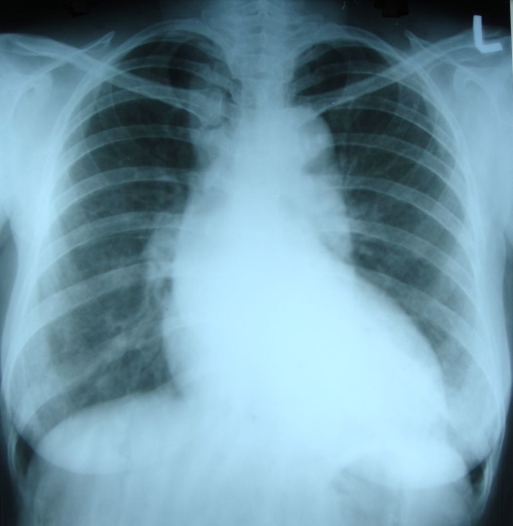
Dilated aorta with unfolding of arch. Ascending aorta is forming the upper part of the right cardiac border and descending aorta the upper part of left cardiac border. The left pulmonary artery is seen as a double density within the descending aortic shadow on the left side. The superior vena caval shadow is seen in the region of the medial end of right clavicle, mainly because the film is rotated towards right as evidenced by the higher distance between the medial end of right clavicle and the spine.
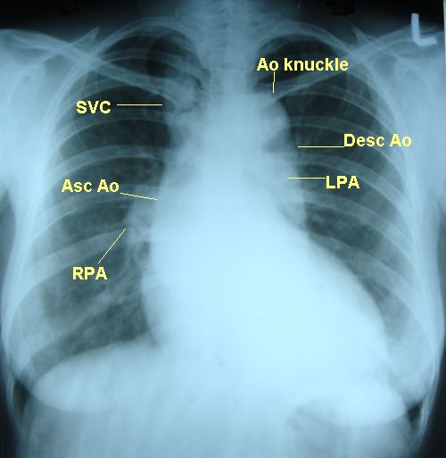
Ao knuckle: aortic knuckle; SVC: superior vena cava; Asc Ao: ascending aorta; Desc Ao: descending aorta; LPA: left pulmonary artery; RPA: right pulmonary artery
Aortic unfolding is often associated with aortic calcification and implies aortic degeneration [1]. Increase in length of ascending aorta with aging is disproportionate to increase in aortic diameter. This appears like a dilated ascending aorta and causes mediastinal widening. Sugawara J et al evaluated arterial lengths using 3-dimensional transverse magnetic resonance imaging in 256 apparently healthy adults with age range 19 to 79 years [2]. They showed that aorta lengthens with age even in healthy persons, primarily due to elongation of ascending aorta.
Lee JW et al evaluated aortic unfolding using non contrast cardiac computed tomography and correlated with age and coronary artery calcium score [3]. It was a retrospective chart review of 219 subjects who had undergone coronary artery calcium scoring during routine health check up. Aortic unfolding was defined by measuring aortic width on non contrast cardiac CT. It was associated with age, body surface area and hypertension. Coronary artery calcium score which is an established surrogate marker of cardiovascular disease was also associated with aortic unfolding. They noted that though aortic unfolding increased progressively with age, it plateaued at 60 years of age. Aortic unfolding index (aortic unfolding indexed for body surface area) plateaued at age ≥70 years. The authors suggested further evaluation of aortic unfolding as potential predictor of cardiovascular risk.
References
- O’Rourke M, Farnsworth A, O’Rourke J. Aortic dimensions and stiffness in normal adults. JACC Cardiovasc Imaging. 2008 Nov;1(6):749-51.
-
Sugawara J, Hayashi K, Yokoi T, Tanaka H. Age-associated elongation of the ascending aorta in adults. JACC Cardiovasc Imaging. 2008 Nov;1(6):739-48.
-
Lee JW, Hur J, Kim YJ, Lee HJ, Nam JE, Kim HY, Hong YJ, Ko SM, Kim TH, Choi BW. Aortic unfolding determined using non-contrast cardiac computed tomography: correlations with age and coronary artery calcium score. PLoS One. 2014 Apr 22;9(4):e95887.
Related Posts
About The Author
Johnson Francis
Former Professor of Cardiology, Calicut Govt. Medical Kozhikode, Kerala, India. Editor-in-Chief, BMH Medical Journal