Doppler in mitral stenosis and regurgitation
Doppler in mitral stenosis and regurgitation
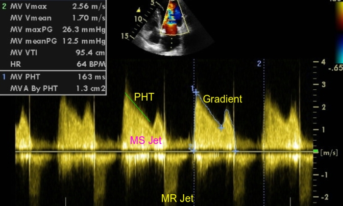
Transmitral Doppler from apical four chamber showing the jet of mitral stenosis (MS Jet) towards the transducer and mitral regurgitation jet (MR Jet) away from the transducer. Estimation of pressure half time (PHT) and transmitral gradient (Gradient) are demonstrated. MV max PG: maximum pressure gradient across the mitral valve; MV mean PG: mean pressure gradient across the mitral valve; MV VTI: mitral velocity time integral; MV PHT: pressure half time of mitral valve; MVA By PHT: mitral valve area by pressure half time.
Mitral valve area by pressure half time
Mitral valve area can be estimated from the pressure half time of the transmitral gradient. Pressure half time is obtained from the deceleration slope of the transmitral E wave in diastole. In mitral stenosis as there is no diastasis, the E merges with the A wave or there is E-A fusion. Green cursor measures the pressure half time of the mitral stenosis jet. Pressure half time is the time taken for the transmitral gradient to fall by half or the transmitral velocity to fall by a factor of square root of two. Pressure half time of two hundred and twenty milliseconds corresponds to a mitral valve area of one square centimeter. Dividing 220 by the measured pressure half time gives the estimated mitral valve area. In this case, the pressure half time was 163 msec and the estimated mitral valve area by pressure half time was 1.3 sq cm.
Smith MD and colleagues have shown that both two dimensional planimetry and pressure half time methods give good estimate of mitral valve area non invasively.1 They found that pressure half time method is superior to two dimensional planimetry to assess mitral valve area in those who have undergone mitral commissurotomy.
It has been shown that associated aortic regurgitation shortens the transmitral pressure half time thereby leading to overestimation of mitral valve area by Doppler echocardiography.2
An earlier study had shown that mitral regurgitation does not significantly change the estimation of mitral valve area by pressure half time method.3
Measurement of transmitral gradient
Transmitral gradient is also measured from the apical four chamber view. The cursor traces the mitral stenosis jet in diastole and the computer program generates the peak and mean diastolic gradient across the mitral valve. Actually the computer calculates the mitral gradient from the transmitral velocity using the simplified Bernoulli equation which states that pressure gradient = 4 V squared.
Mitral regurgitation jet by Doppler
The mitral regurgitation jet (MR Jet) by Doppler is seen away from the transducer (below the baseline). It is an incomplete jet suggesting that mitral regurgitation is only mild.
References
- Smith MD, Handshoe R, Handshoe S, Kwan OL, DeMaria AN. Comparative accuracy of two-dimensional echocardiography and Doppler pressure half-time methods in assessing severity of mitral stenosis in patients with and without prior commissurotomy. Circulation. 1986 Jan;73(1):100-7.
- Flachskampf FA, Weyman AE, Gillam L, Liu CM, Abascal VM, Thomas JD. Aortic regurgitation shortens Doppler pressure half-time in mitral stenosis: clinical evidence, in vitro simulation and theoretic analysis.J Am Coll Cardiol. 1990 Aug;16(2):396-404.
- Hatle L, Angelsen B, Tromsdal A. Noninvasive assessment of atrioventricular pressure half-time by Doppler ultrasound. Circulation. 1979 Nov;60(5):1096-104.
Related Posts


About The Author
Johnson Francis
Former Professor of Cardiology, Calicut Govt. Medical Kozhikode, Kerala, India. Editor-in-Chief, BMH Medical Journal