Double atrial shadow and prominent upper lobe vessels
Double atrial shadow and prominent upper lobe vessels
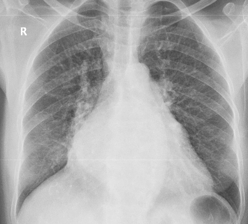
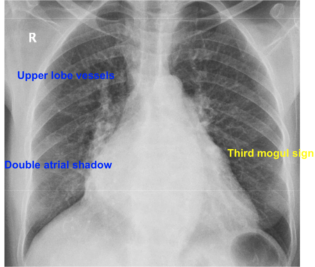
X-ray chest PA view showing prominent double atrial shadow (double atrial contour, shadow in shadow, double density [1]) to the right of the spine, suggestive of left atrial enlargement. This pattern is typically seen in long standing mitral stenosis with or without regurgitation. Double atrial shadow has also been described in a case of cor triatriatum with myxomatous mitral valve degeneration producing severe mitral regurgitation [2]. Cor triatriatum can masquerade as mitral stenosis.
Here the left atrial shadow reaches up to the upper part of the right heart border. It may be noted that normally right atrium forms the right heart border and left atrium does not reach the right heart border. Left atrium reaches the right border when it is significantly enlarged. Grossly enlarged left atrium when it is aneurysmal, may even bulge further beyond the right atrium on the right heart border in PA view [3]. Such gross left atrial enlargement is likely in long standing severe mitral regurgitation.
Another sign of left atrial enlargement on chest X-ray is elevation of left bronchus with widening of the tracheal bifurcation carinal angle [4].
Right border of the heart is significantly shifted to the right, indicating associated right atrial enlargement. Left atrial appendage is seen as a bulge along the middle region of left heart border (third mogul sign) [5]. Upper lobe vessels are quite prominent (inverted moustache sign, antler sign [6], cephalization), indicating pulmonary venous hypertension. All together the findings on the chest X-ray indicate severe mitral stenosis. While evaluating chest X-ray in such cases of long standing mitral stenosis, close scrutiny for visible calcification of mitral valve is needed. Milder degrees of calcification of mitral valve is better appreciated on fluoroscopy.
References
- Higgins CB, Reinke RT, Jones NE, Broderick T. Left atrial dimension on the frontal thoracic radiograph: a method for assessing left atrial enlargement. AJR Am J Roentgenol. 1978 Feb;130(2):251-5.
- Wong CK, Leung WH, Cheng CH, Lau CP, Cheung DL. Myxomatous mitral valve degeneration complicating asymptomatic cor triatriatum. Clin Cardiol. 1989 Jan;12(1):48-50.
- Le Roux BT, Gotsman MS. Giant left atrium. Thorax. 1970 Mar;25(2):190-8.
- Karabulut N. CT assessment of tracheal carinal angle and its determinants. Br J Radiol. 2005 Sep;78(933):787-90.
- Gorospe L, Fernández-Méndez MÁ, Ayala-Carbonero AM, García-Poza J, González-Gordaliza C. Ortner’s Syndrome Secondary to a Huge Left Atrium. Ann Thorac Surg. 2015 Aug;100(2):732.
- Han J, Xiang H, Ridley WE, Ridley LJ. Antler sign: Pulmonary venous hypertension. J Med Imaging Radiat Oncol. 2018 Oct;62 Suppl 1:13. A chest X-ray with overlay of the picture of an antler is available at the journal site.
Related Posts

About The Author
Johnson Francis
Former Professor of Cardiology, Calicut Govt. Medical Kozhikode, Kerala, India. Editor-in-Chief, BMH Medical Journal
Thanks for a very good x ray.
Inverted moustache sign, Grade 2 LA enlargement with double atrial shadow, splaying of carina (>90 degrees), third mogul sign of LA appendage, elevation and narrowing of left main bronchus and probable pulmonary bay obliteration can be seen.