ECG filter settings and detection of pacemaker spikes
ECG filter settings and detection of pacemaker spikes
In modern day digital ECGs, ECG filter settings can beautify the tracings, but vital information can be filtered out, as in this case, which is quite common. In the illustration below, 0.08 Hz is the high pass filter, meaning that the ECG amplifier passess all frequencies above that limit. 40 Hz is the low pass filter, indicating that all frequencies below that can pass through.
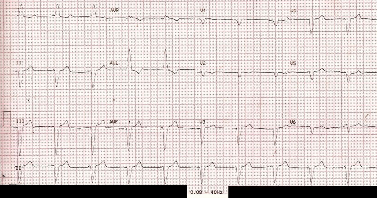
Pacemaker spike (pacemaker artefact, pacemaker stimulus) being a high frequency signal, is effectively filtered out by the above filter setting. Hence the above ECG is likely to be diagnosed as left bundle branch block (LBBB) at one look, though we are missing the P waves before each QRS complex which would be expected in a simple LBBB with sinus rhythm.
 ECG with filter range 0.08 – 150 Hz – pacemaker spikes visible
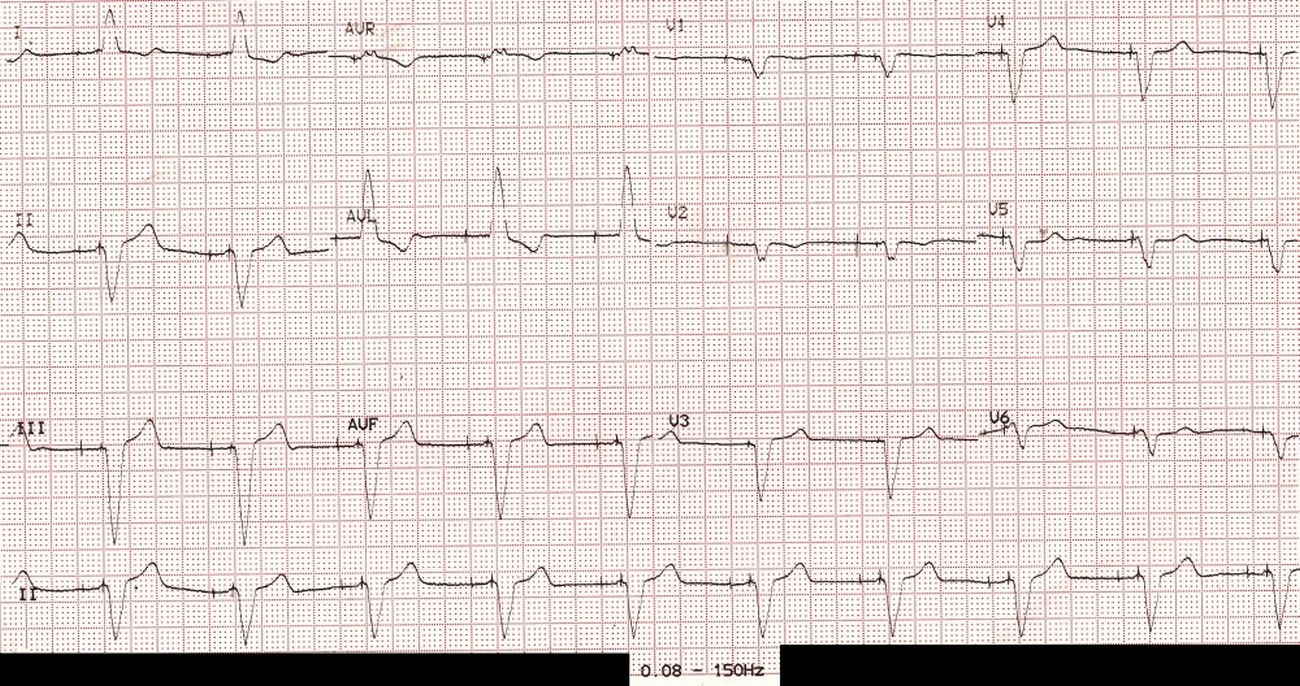
The pacemaker spikes are easily seen when the same ECG is repeated with a change in the low pass filter to 150 Hz. In fact both atrial and ventricular pacing spikes are seen before each QRS complex, with an interval in between. Hence this person has a functioning dual chamber pacemaker in situ. The paced P waves are of low amplitude and hardly visible (or it could be lack of atrial response to atrial pacing – atrial capture failure). This highlights the importance of appropriate ECG filter settings for each case.
The high pass filter is meant to filter out baseline fluctuations due to respiration and other low frequency signals, while low pass filter avoids muscle potential interference (EMG artefact). In addition, most ECG machines have a notch filter at 50 Hz to specifically filter out line voltage interference.
About The Author
Johnson Francis
Former Professor of Cardiology, Calicut Govt. Medical Kozhikode, Kerala, India. Editor-in-Chief, BMH Medical Journal