Old anterior wall myocardial infarction and left atrial overload
ECG Quiz 14
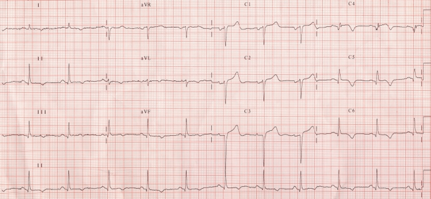
Left atrial overload is seen as prominent negative component of P wave in lead V1. There is poor progression of R waves in anterior leads with QS complex in lead V3 and qr in V4. T waves are inverted in leads I, II, aVl and V4 to V6. ST segment is almost isoelectric. These findings together indicate that the myocardial infarction was remote. If the ST segment elevation is persisting for a long period after myocardial infarction, it is considered as a feature of left ventricular aneurysm.
Related Posts

About The Author
Johnson Francis
Former Professor of Cardiology, Calicut Govt. Medical Kozhikode, Kerala, India. Editor-in-Chief, BMH Medical Journal