Lateral Wall Ischemia and ?LVH
Lateral Wall Ischemia and ?LVH
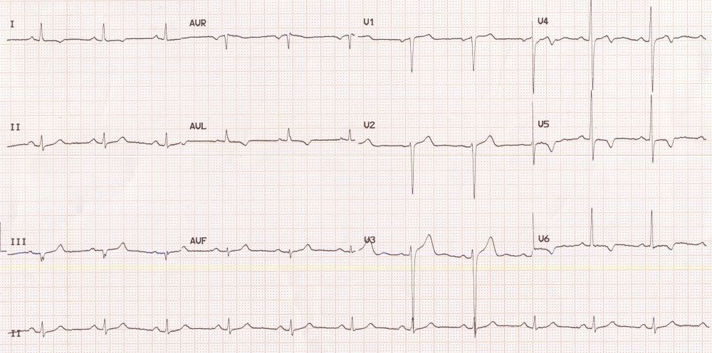
ECG shows T wave inversion in lateral leads I, aVl, V5 and V6, suggestive of lateral wall ischemia. Deep S waves in V2 and tall R waves in V5 give an indication of left ventricular hypertrophy, though the S wave in V1 is not that deep and the R wave in V6 is not that tall. Ventricular rate is on the lower side indicating the possible use of drugs which suppress the sinus node. This combination could occur in hypertensive individuals with coronary artery disease being treated with beta blockers or nondihydropyridine group of calcium channel blockers. Lower heart rate can also be due to intrinsic sinus node dysfunction.
Related Posts

About The Author
Johnson Francis
Former Professor of Cardiology, Calicut Govt. Medical Kozhikode, Kerala, India. Editor-in-Chief, BMH Medical Journal